3 min read 5

Anatomy on Radiographs: Pantomographs Part 1

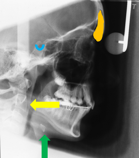
Ok, I am back and now onto pantomographs. Since these images are larger and will take up much more space I have decided to break them down into 2 parts.  I also will have several anatomy identified on each radiograph as well.  Here we go. Mandible *The mandibular condyle (two…
2 min read 7

Anatomy on Radiographs: Intraoral Radiographs Part 2

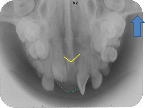
This is part 2 (posterior) of anatomy on intraoral radiographs. Mandible The mental foramen  appears as a round to oval radiolucent area near the apex of the second premolar. The inferior alveolar nerve canal (mandibular canal) appears as radiolucent band with two thin radiopaque lines running parallel to each other…
1 min read 0

Case of the Week: Mucous retention pseudocyst

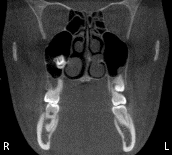
This week I have a case of a mucous retention pseudocyst on a pantomograph.  Mucous retention pseudocysts are incidental findings that do not require treatment.  This can occur on any surface of the sinus but is most commonly seen on the floor.  Note the rounded radiopaque dome in the left…
1 min read 0

Starting new posts and updates to pages

I will be starting to add to this blog more frequently and also creating pages to complete this source of radiographs for all dental professionals.  I will be posting on other oral radiology websites, radiographic interpretation, showing clinical cases, and other fun tips or answers to common oral radiology questions.…