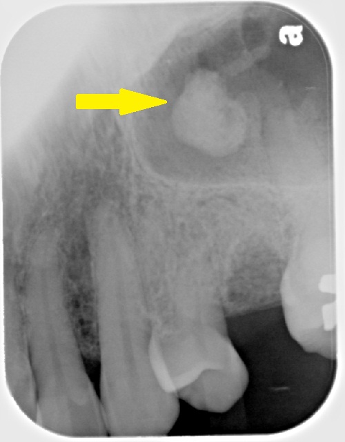
Definition: A calcification in the maxillary sinus. This calcification may be of long standing mucous or foreign bodies, including tooth fragments.
Radiographic Features:
Location: Maxillary sinuses.
Edge: Well-defined, smooth or irregular outline.
Shape: Round, ovoid.
Internal: Radiopaque, may have a ‘laminated’ appearance with radiopaque and radiolucent bands evident due to continued laying down of calcium salts. (This looks similar to layers of an onion.)
Other: None
Number: May be single of multiple.
TIP: Evaluate that the radiopaque area does not appear to be attached to a border of the maxillary sinus. If it appears to be attached to a border on multiple images, an antral exostoses/antral projection should be considered.
(click image to enlarge)
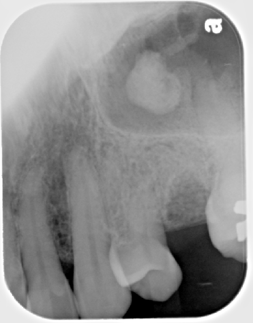
Antrolith
(arrow pointing to well-defined radiopaque area not attached to the border of the maxillary sinus)
Antrolith
(without arrow)
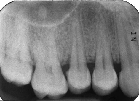
Antrolith
(superior to the maxillary right first molar – # 3)
Case of the week


