Here is the first section when creating a radiology report with the main areas I would cover with students along with links to pages for additional information and examples.
First, is to identify teeth present. There are many ways to go about this. Below are examples of a table I used to show which teeth were present along with a few identifiers. The line in the middle is the occlusal plane and teeth numbers are moved as needed. (These are in the Universal Numbering System, so 1-32 for permanent teeth and A-T for primary teeth.)
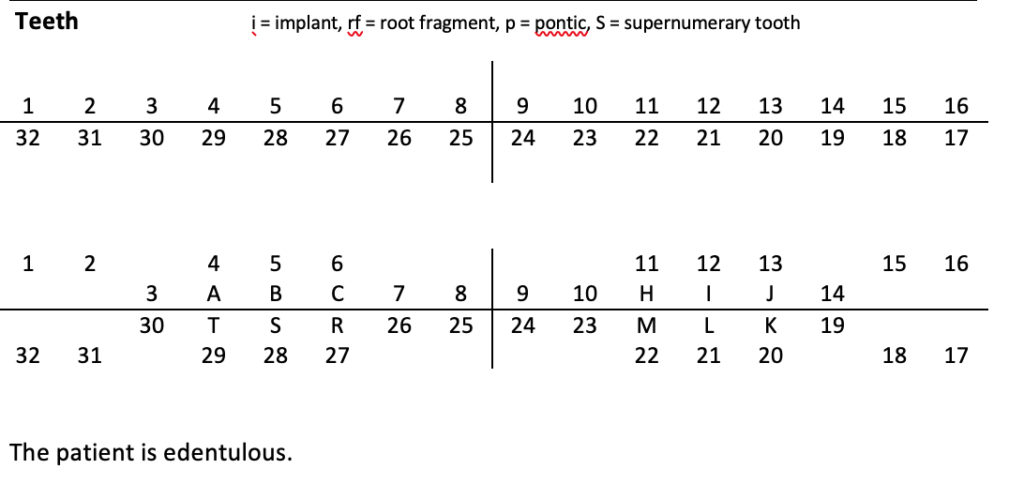
I’ve seen others list out the teeth present such as 2-15 and 18-31. You can also include more about dental treatment present such as endodontically treated teeth and teeth with restorations. If a patient is edentulous, I end the section with that as there are no teeth to evaluate. 🙂
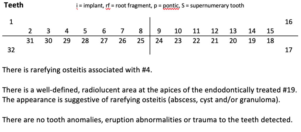
Second is periapical inflammatory lesions such as rarefying osteitis, sclerosing osteitis, and/or a widened periodontal ligament space. If there are no periapical inflammatory lesions, I include a negative statement such as ‘no periapical inflammatory lesions detected’.
Third is all about tooth anomalies and eruption abnormalities. The tooth anomalies include both internal findings (such as pulp stones, internal resorption, etc.) and external findings (such as external resorption, dilaceration, talon cusp, etc.). The eruption abnormalities include findings such as ankylosis, ectopic eruption, etc. Again if nothing to comment on, include a negative statement.
Last is all about trauma to the teeth. This includes root fractures, abnormal bone healing variants, etc.
Here’s a sample of how one completed looks.
The next section is caries coming next week.
Thanks and enjoy!

One thought on “Teeth: Creating a systematic radiology report for 2D radiographs”
Comments are closed.