Definition: An antral exostoses (antral projection) is an exostoses (outward growth of bone) into the maxillary sinuses.
Radiographic Features:
Location: Frequently seen on the floor of the maxillary sinus.
Edge: Well-defined, attached to a border of the maxillary sinus.
Shape: Ovoid, round, appears ‘mushroom’ shaped on 3-D images.
Internal: Radiopaque, same radiopacity as bone.
Other: None.
Number: Usually single, but may be multiple.
TIP: When viewing an antral exostoses (antral projection) on a periapical radiograph evaluate if the radiopaque area is attached to the border of the maxillary sinus. If the radiopaque area is not attached to the border, an antrolith should be considered.
(click image to enlarge)
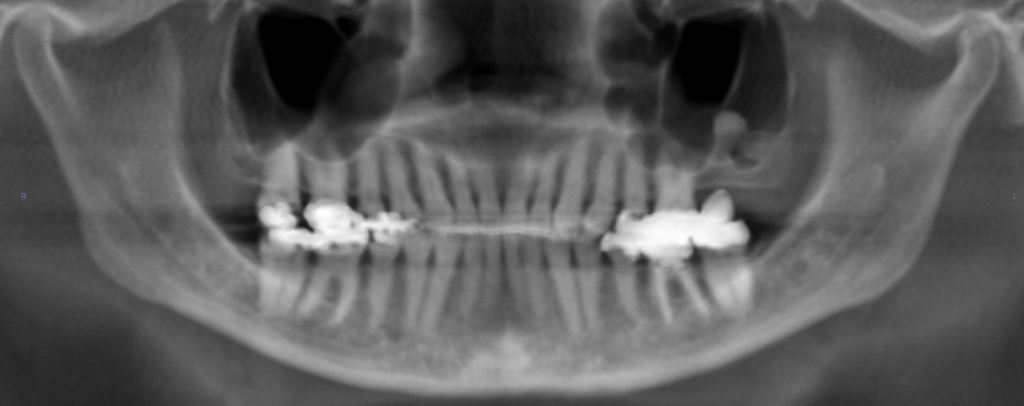
Antral Exostoses/Antral Projection
(arrow – note how the radiopaque area appears to stay attached to the border on both images)
 Antral Exostoses/Antral Projection
(without arrow)
 Antral Exostoses/Antral Projection
 Antral Exostoses/Antral Projection
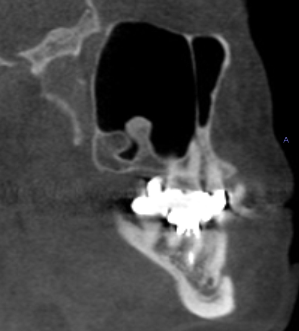
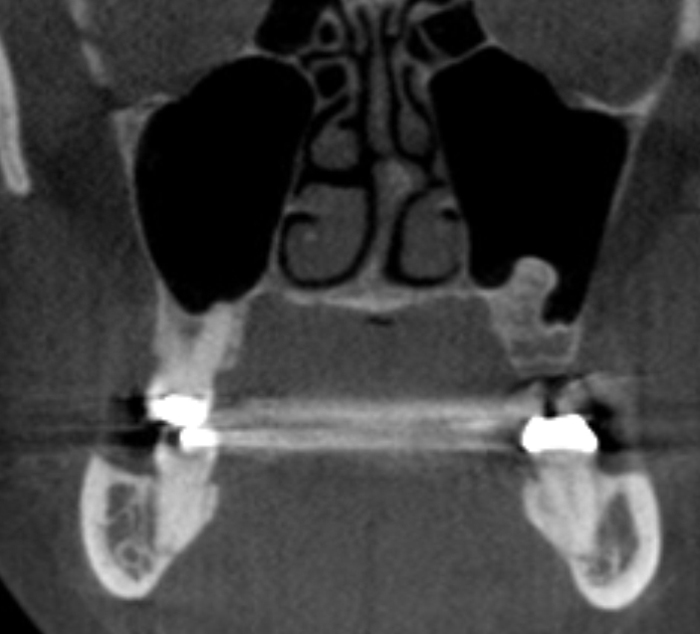
(Cone Beam CT – Reconstructed pantomograph, coronal and sagittal views showing 2 antral exostoses/antral projections in the left maxillary sinus)