1 min read 0

Lateral Cephalometric Skull Anatomy – Part VI

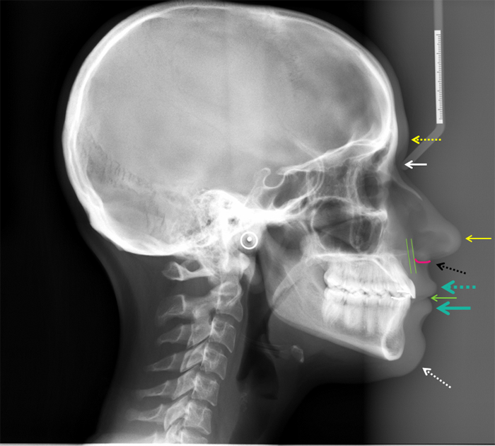
This is the final post and it will cover soft tissue anatomy seen on lateral cephalometric skull radiographs. Glabella (yellow dotted arrow) Nasion (white solid arrow) Pronasale (yellow solid arrow) Subnasale (black dotted arrow) Labrale superius (turquoise dotted arrow) Stomion (green solid arrow) Labrale inferius (turquoise solid arrow) Gnathion (white…
1 min read 0

Lateral Cephalometric Skull Anatomy – Part V

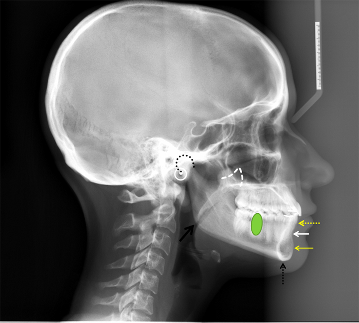
Today is all about the mandible. Condyle (black curved dotted line) Coronoid process (white triangular dotted line) Mandibular first molar (green oval) Infradentale (yellow dotted arrow) – Superior facial bone height near the level of the cemento-enamel junction of the mandibular incisors. B point (white solid arrow) – The posteriormost…
1 min read 0

Lateral Cephalometric Skull Anatomy – Part IV

This post will be all about the maxilla. Anterior nasal spine (yellow arrow) – anterior-most point where right and left maxilla meet on the midline forming a protruding bone spicule. A point (white arrow) – the deepest point between the anterior nasal spine and prosthion. Prosthion (yellow dotted arrow) –…
1 min read 0

Lateral Cephalometric Skull Anatomy – Part III

Now onto the temporal region. Zygomatic Arch (green area) – Made up by the zygomatic bone and zygomatic process of the temporal bone. Mandibular Notch (blue line) – Notch between the condyle and coronoid process of the mandible. Posterior margin of the ramus (white line) – Just as the name…
1 min read 8

Lateral Cephalometric Skull Anatomy – Part II

Onto part 2 of lateral cephalometric skull anatomy frequently used in orthodontics. Again identified on a dry skull first followed by the radiograph. Orbitale (black star) – The lowest point of the orbital margin (also the anterior point for the Frankfort horizontal plane). Key ridge (green heart) – The inferior…