This week will be covering the first 2 criteria for an ideal pantomograph and what happens to the final image when they are not met.
1. Patient has anterior teeth in notches on bite block.
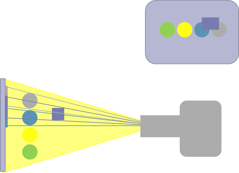
- Error: Patient positioned too far anteriorly or posteriorly in the unit creating minification (too far anteriorly) of magnification (too far posteriorly) of the anterior teeth.
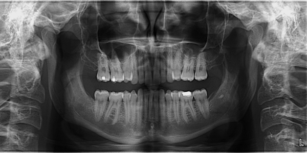
Note the smaller than normal appearance of the anterior teeth
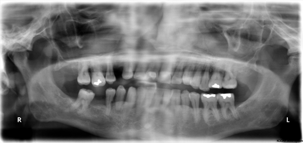
Note the larger than normal appearance of the anterior teeth
- Correction: Ensure patient has teeth in notches of bite block.
2. Patient has midline (anterior and posterior of head) aligned with sagittal midline indicator light creating symmetry between the left and right side of the patient.
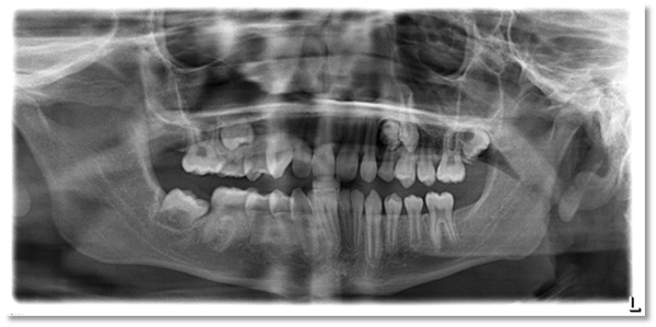
- Error: Patient’s midline is not aligned with the sagittal midline indicator light creating asymmetry between the left and right sides.
Note the anatomy (including jaws and teeth) on right side is larger compared to the left side.
- Correction: Ensure patient midline (both anteriorly and posteriorly) is aligned with the sagittal midline indicator light.
In two weeks: Criteria 3 and 4 (Next week is How was that missed)
If you have any questions, comments or good examples of errors regarding these two specific criteria, please let me know. Thanks and enjoy!
SPONSOR
One thought on “Radiographic Quality Evaluation: Bad Pantomographs (Part 1)”
Comments are closed.