Now onto other structures visible on a pantomograph.
Midface
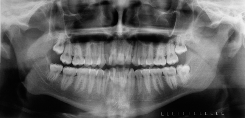
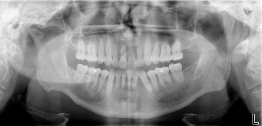
The orbital rim (green dots) appears as a thick semi –circular radiopaque band superior to the maxillary sinuses.
The pterygomaxillary fissure (blue area) appears as an inverted teardrop shaped radiolucent area with a thin radiopaque border. It is directly lateral to the maxillary sinus.
The zygomatic bone (yellow line)appears as a radiopaque area distal to the zygomatic process of the maxilla. It is superimposed over the distal aspect of the maxillary sinus and pterygomaxillary fisure.
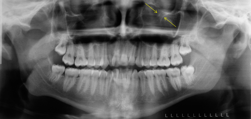
The infraorbital canal (yellow arrows) appears a radiolucent band with two thin radiopaque lines running parallel to each other as the borders of the canal. It is angled infero-medially and superimposed over the orbital rim.
Other
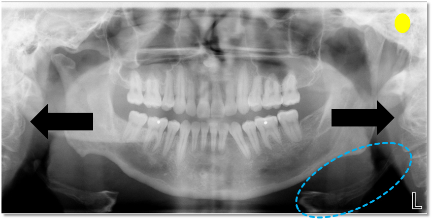
The external auditory meatus (yellow circle) appears as a round to ovoid radiolucent area lateral to the glenoid fossa and condyle.
The cervical spine (black arrows) appears radiopaque and is seen on the lateral aspect of the pantomograph. It is not seen on all patients and visualization is dependent on patient positioning and size.
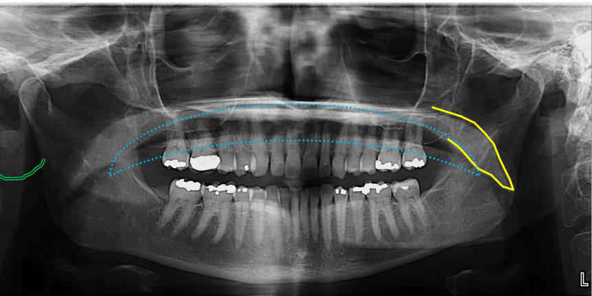
The hyoid (blue dotted circle) appears radiopaque inferior to and/or superimposed over the inferior border of the mandible.
The ear lobe (double green curved lines) appears as a radiopaque area lateral to and/or superimposed over the ramus of the mandible.
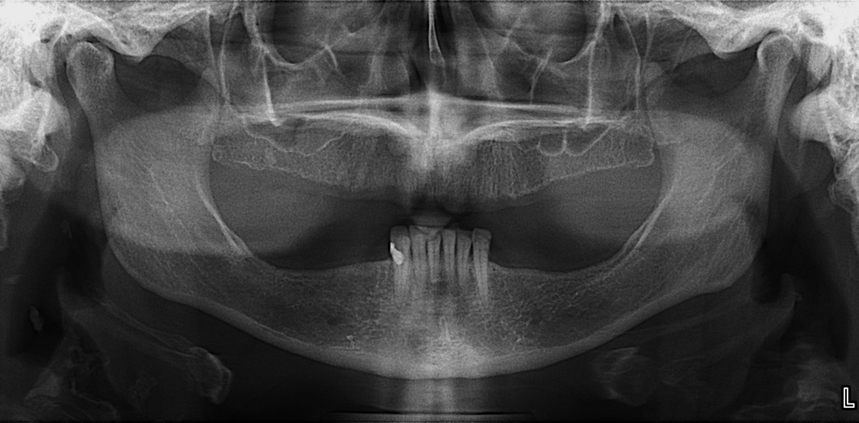
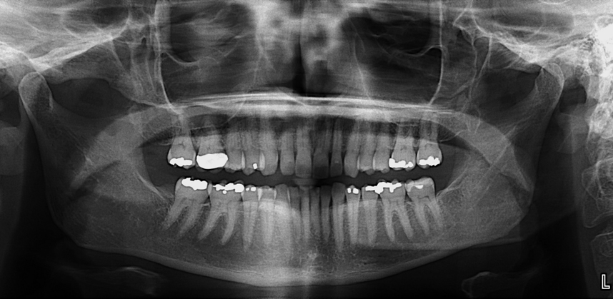
The oropharynx (blue dotted line) appears as a radiolucent band superimposed over the roots of the maxillary teeth directly inferior to the floor of the nasal cavity. This is only seen when the patient does not position their tongue to the roof of the mouth during the exposure.
The soft palate (yellow line) appears as a radiopaque mass coming off the floor of the nasal cavity. This is more readily seen when the patient does not have their tongue on the roof of the mouth during exposure.
Next = Extraoral Radiographs or Occlusals (I haven’t decided yet which one so it’ll be a little surprise next week)
I hope you find this second post on pantomograph anatomy informative. While I didn’t cover all the anatomy visible, these are the more commonly seen entities. If you have any questions, please let me know.
Enjoy!

Dr, Could you please indicate the airspaces in the image- palatoglossal, nasopharyngeal and glossopharyngeal?
Thanks
I will create some Anatomy Monday posts in the coming weeks showing these areas individually. Thanks for the request. 🙂
Thank you so much. That would be really helpful 😉
Would you like answer my question what type of image do you need for TMJ?
Are you asking what type of radiographs are recommended for imaging of the TMJ area? Thanks.
Yes. I mean in which kind of x ray we can have better view of TMJ?
Pantomographs provide a basic but rotated view of the condyle and fossa. 3D imaging is recommended for further evaluation such as a cone beam CT, spiral CT or MRI if investigating the soft tissues.
I got it. Thank you so much!
These are great! Thank you!
🙂