zygomatic bone


Anatomy Monday: Zygomatic Bone

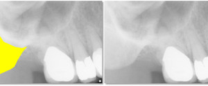
Now that you know how to identify the zygomatic process of the maxilla it’s very easy to find the zygomatic bone.  The zygomatic bone presents as a well-defined radiopaque area directly lateral/distal to the zygomatic process of the maxilla. If you have any questions or comments about the zygomatic bone, […]


Anatomy on Radiographs: Pantomographs Part 2 10

Now onto other structures visible on a pantomograph. Midface The orbital rim (green dots) appears as a thick semi –circular radiopaque band superior to the maxillary sinuses. The pterygomaxillary fissure (blue area) appears as an inverted teardrop shaped radiolucent area with a thin radiopaque border. It is directly lateral to […]


Anatomy on Radiographs: Intraoral Radiographs Part 2 7

This is part 2 (posterior) of anatomy on intraoral radiographs. Mandible The mental foramen  appears as a round to oval radiolucent area near the apex of the second premolar. The inferior alveolar nerve canal (mandibular canal) appears as radiolucent band with two thin radiopaque lines running parallel to each other […]