Happy New Year! Starting off with a fun and rare case that I don’t see too often.
This week I have an interesting case of two different lesions (florid cemento-osseous dysplasia and simple bone cyst) that have been reported together. The combination of the two were first reported in 1976. This is case combination is a rarity. I’m sharing this case to emphasis that importance of thorough investigation of florid cemento-osseous dysplasia along with continual monitoring. I’ll go over the two radiographic appearances of each lesion using my LESION acryonym.
Florid Cemento-osseous dypslasia
- Location = involves two quadrants or four quadrants (mandible / maxilla or both).
- Edge = well-defined.
- Shape = no identifiable shape.
- Internal aspect = Stage 1 – purely radiolucent, Stage 2 – mixed radiolucent/radiopaque, Stage 3 – purely radiopaque (+/- radiolucent rim visible).
- Other = +/- thinning of cortical plates.
- Number = one in one jaw, two in both jaws.
- Location = more common in the mandibular premolar/molar region.
- Edge = well-defined, thin corticated border.
- Shape = scallops between roots, ‘hydraulic’ appearance, +/- round/ovoid.
- Internal aspect = radiolucent.
- Other = +/- displace adjacent teeth over time, +/- thinning of cortical plates.
- Number = single.
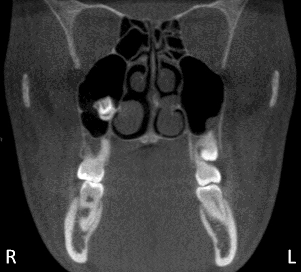
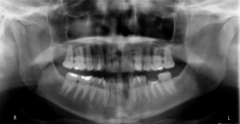
This case is from a cone beam CT with videos. The simple bone cyst on this case is in the posterior right mandible.