Today I thought I’d go with a little refresher of foramina visible on intraoral radiographs specifically periapical radiographs. There will be a total of 4 I’m covering, 2 each in the maxilla and mandible. So onto the maxilla first.
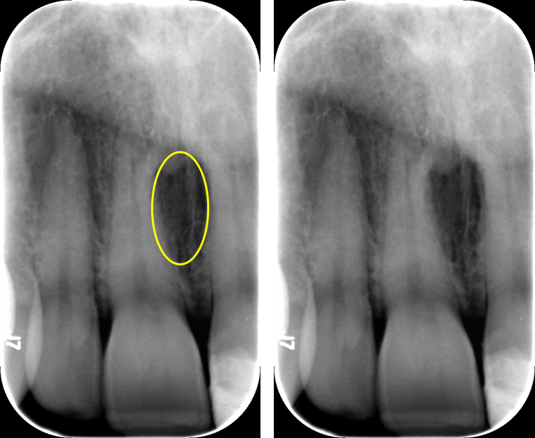
Incisive Foramen
The incisive foramen is the inferior opening of the nasopalatine canal (incisive canal). It is opens between the roots of the maxillary central incisors on the lingual. It will appear as a round to ovoid radiolucent area between the roots.

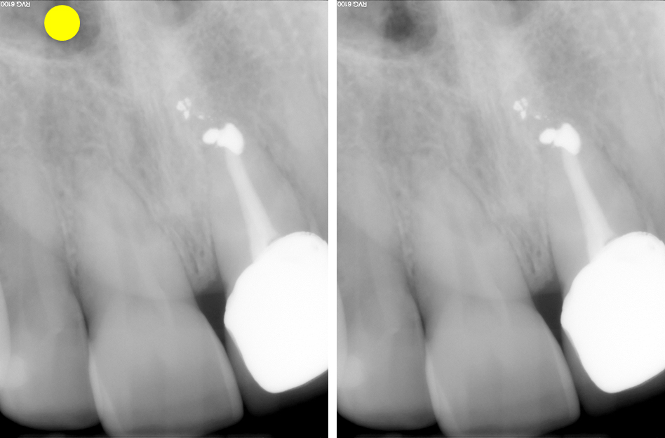
Superior Foramina of the Nasopalatine Canal
The superior foramina of the nasopalatine canal is exactly what the names states. It opens onto the floor of the nasal cavity. There are two of these foramen as the canal splits when going superiorly with one opening in each side of the nasal cavity. It appears as round radiolucent area just lateral to the nasal septum and superior to the floor of the nasal cavity.

Now, onto the mandible.
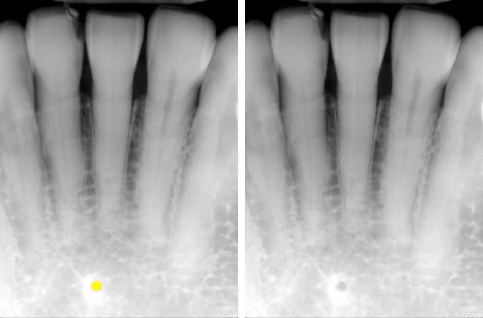
Lingual Foramen
The lingual foramen is a term that applies to any foramen that opens on the lingual aspect of the mandible but for purposes of intraoral imaging the term is used when referring to the foramen or foramina that open in the midline of the mandible on the lingual side. There can be up to 3 foramina in the midline on some patients. The multiple foramina are more commonly seen on cone beam CT imaging. It appears as a small very round (similar to a hole punch) radiolucent area in the midline of the mandible inferior to the apices of the central incisors.

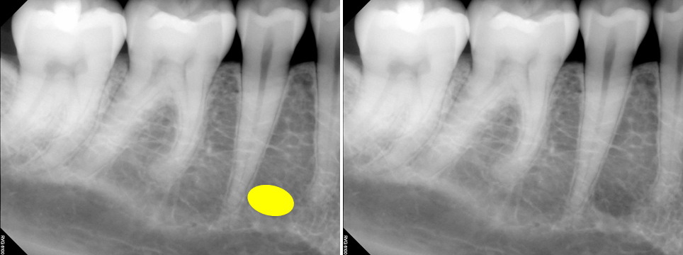
Mental Foramen
The mental foramen is an opening of the inferior alveolar nerve canal (mandibular canal). It opens on the facial of the mandible near the apex of the second premolar. It appears as a round to ovoid radiolucent area inferior to or superimposed over the apex of the second premolar. It can also been seen mesially up to the apex of the first premolar or distally to the mesial apex of the first molar.

TIP: For the incisive foramen and mental foramen, if there is a question of the area being possible pathosis associated with the adjacent tooth look for the periodontal ligament space. If the periodontal ligament space is intact around the root and through the radiolucent area, it is likely a foramen and not pathosis associated with the tooth.
If you have any questions or comments, please leave them below. Thanks and enjoy!

Pingback: Anatomy Monday: Canals on Intraoral Radiographs – Dr. G's Toothpix