1 min read
2
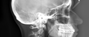
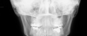
Radiographic Technique: PosteroAnterior (PA) Skull Radiograph
This week is a less commonly made extraoral radiograph but still occasionally seen in dentistry. Here is how to position a patient for this radiograph. 1. Place the coronal plane of the patient parallel with the image receptor. If there is a craniostat to help position this will be GENTLY…