1 min read 0

Case of the Week: Mucous Retention Pseudocyst

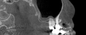
This week I have a case of a mucous retention pseudocyst on a pantomograph. Mucous retention pseudocysts are incidental findings that do not require treatment. They are most commonly found in the maxillary sinuses followed by the sphenoid sinuses. Note the rounded radiopaque dome in the right maxillary sinus. If…
1 min read 0

Educational Video: Sinusitis

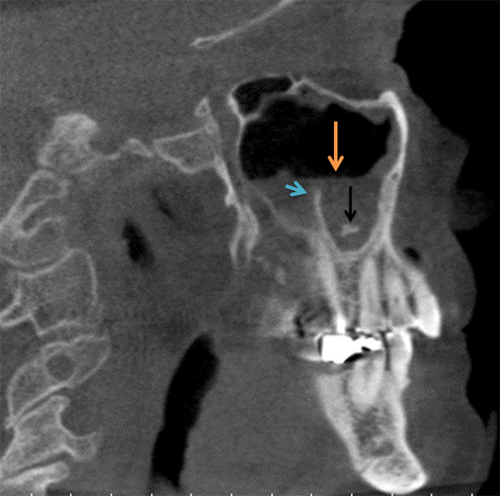
This week I have a case of sinusitis on cone beam CT along with another educational video. 🙂 On the cone beam CT images note the radiopaque band that follows the floor and the posterior border on the sagittal view and the floor and medial border of the left maxillary…
1 min read 4

Case of the week: Antrolith and Sinusitis

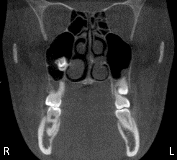
This weeks case has a few things going on in one of the maxillary sinuses.  There are three different findings in the left maxillary sinus: an antrolith, sinusitis, and a maxillary sinus septum. An antrolith is a calcification within the maxillary sinuses. Sinusitis is thickening of the soft tissue of…
2 min read 2

Case of the week: Third Molar in Maxillary Sinus

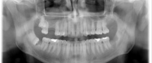
This weeks case topic is an interesting finding of a maxillary third molar developing in the maxillary sinus adjacent to a supernumerary tooth on a CBCT scan.  Interpretation: This case shows a mixed radiolucent/radiopaque entity in the right maxillary sinus.  The radiopacity is that of tooth structure.  The appearance is…
2 min read 0

Anatomy on Radiographs: Occlusal Radiographs

This is the last post I have planned for anatomy and it will be on occlusal radiographs. Mandible Short and sweet here with only one anatomical landmark readily visible. The genial tubercles (yellow arrow) appear as a small radiopaque mass(es) extending off the lingual aspect of the mandible. Maxilla The nasal…