5 min read 0

Anatomy Monday: Anatomy on Mandibular Periapical Radiographs

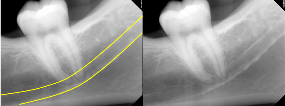
It’s been a while since I’ve updated anatomy seen on intraoral radiographs so I’m continuing with all the anatomy visible on mandibular periapical radiographs. Some of these were seen in the recent posts on canals and foramina but this will be the one stop spot for everything mandibular periapical radiographs.…
3 min read 1

Anatomy Monday: Foramina on Intraoral Radiographs

Today I thought I’d go with a little refresher of foramina visible on intraoral radiographs specifically periapical radiographs.  There will be a total of 4 I’m covering, 2 each in the maxilla and mandible.  So onto the maxilla first. Incisive Foramen The incisive foramen is the inferior opening of the…
1 min read 2

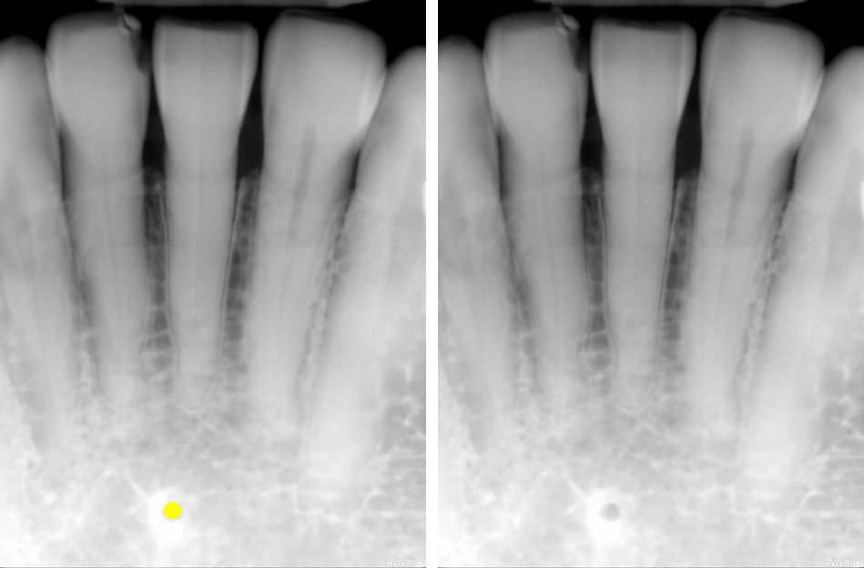
Anatomy Monday: Lingual Foramen (mandible)

It’s another foramen in the mandible.  While there are multiple lingual foramina in the mandible, the most commonly seen one is in the anterior mandible on the midline.  It appears as a round radiolucent entity with a thin to thick corticated border.  It may be single or multiple (up to…
1 min read 4

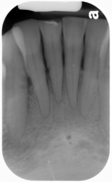
Case of the week: Double Lingual Foramina

This weeks case is an interesting variant of normal anatomy – two lingual foramina seen on a periapical radiograph.  Some patients can have up to three lingual foramina. Note the two well-defined, circular radiolucent entities inferior to the mandibular central incisors.  The superior one is larger than the inferior one.…
3 min read 5

Anatomy on Radiographs: Pantomographs Part 1

Ok, I am back and now onto pantomographs. Since these images are larger and will take up much more space I have decided to break them down into 2 parts.  I also will have several anatomy identified on each radiograph as well.  Here we go. Mandible *The mandibular condyle (two…