1 min read 0

Case of the week: Internal Resorption on CBCT

This weeks case is a severe example of internal resorption as seen on cone beam CT.  Internal resorption presents as enlargement of the pulp chamber and/or root canal space. Reconstructed pantomograph (Metal in left  anterior mandible is due to previous reconstruction and mandibular fracture) Cross-sectional view (Note enlarged root canal…
2 min read 2

Case of the week: Third Molar in Maxillary Sinus

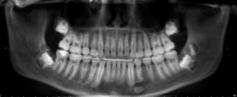
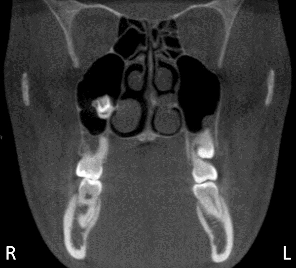
This weeks case topic is an interesting finding of a maxillary third molar developing in the maxillary sinus adjacent to a supernumerary tooth on a CBCT scan.  Interpretation: This case shows a mixed radiolucent/radiopaque entity in the right maxillary sinus.  The radiopacity is that of tooth structure.  The appearance is…
2 min read 0

Case of the week: Dentin Dysplasia Type II

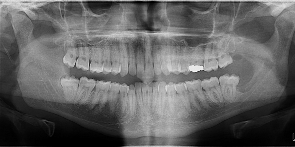
Due to ensuring this site in no way violates HIPPA and pleases the lawyers, I am changing the series title of ‘This week in the clinic’ to ‘Case of the week’ to ensure there is no confusion that the case presented was possibly seen in the clinic that specific week…
1 min read 0

Case of the Week: Blunderbuss apex

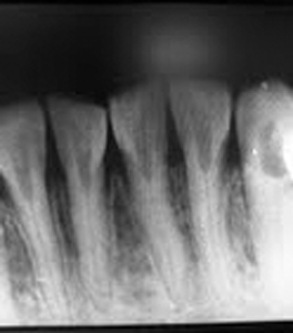
This week I wanted to show normal anatomy of a developing tooth; the blunderbuss apex.  A blunderbuss apex is an open apex of a developing tooth. It is named blunderbuss due to the similar appearance to a blunderbuss rifle.  An open or blunderbuss apex appears as pyramidal radiolucent area.  This…