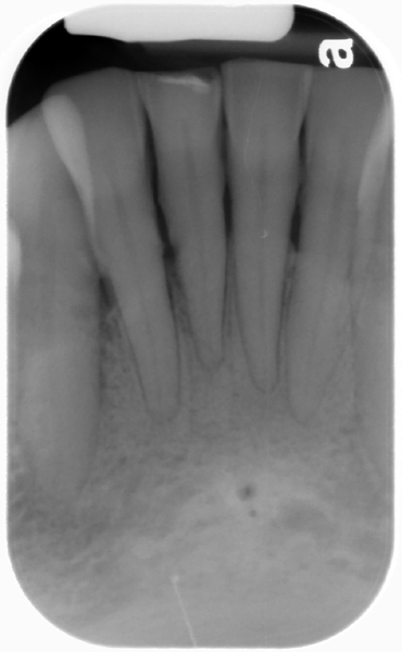
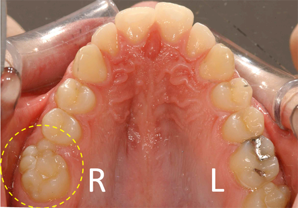
1 min read 4

Case of the week: Double Lingual Foramina

This weeks case is an interesting variant of normal anatomy – two lingual foramina seen on a periapical radiograph.  Some patients can have up to three lingual foramina. Note the two well-defined, circular radiolucent entities inferior to the mandibular central incisors.  The superior one is larger than the inferior one.…
1 min read 4

Case of the week: Chronic Apical Periodontitis

This week is a follow up to last week with a case of chronic apical periodontitis.  The primary difference of chronic apical periodontitis versus acute apical periodontitis is the presence of sclerotic bone formation.  This sclerotic bone formation (sometimes referred to as sclerosing osteitis) may present in one of three…
2 min read 0

Case of the week: Apical Periodontitis (Apical Rarefying Osteitis)

This week’s case is from a request to see cases of apical periodontitis and chronic apical periodontitis (coming next week).  A good definition I found of apical periodontitis is “a spectrum of diseases that occur around the tooth apex, including periapical granuloma, periapical abscess, and periapical (radicular) cyst” (Scheinfeld MH,…
1 min read 4

Case of the week: Antrolith and Sinusitis

This weeks case has a few things going on in one of the maxillary sinuses.  There are three different findings in the left maxillary sinus: an antrolith, sinusitis, and a maxillary sinus septum. An antrolith is a calcification within the maxillary sinuses. Sinusitis is thickening of the soft tissue of…
1 min read 4

Case of the week: Enamel Hypoplasia

This week I have a case of enamel hypoplasia on the maxillary central incisors.  I have no information on the history of this case and any possible causes. If you have some ideas, please feel free to share in the comments. Note the wavy radiolucent lines in the middle portion…