1 min read 0

Anatomy Monday: Epiglottis (soft tissue)

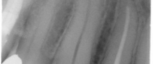
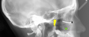
This week I wanted to show off something not commonly looked at or for on pantomographs; the epiglottis. The epiglottis will appear near the inferior aspect of the radiograph as a curved radiopaque entity. It may be superimposed over the hyoid or inferior border of the mandible.  If the airway…
1 min read 2

Case of the week: Horizontal root fracture

This week I have a case of a horizontal root fracture that is tricky to see on the initial radiograph but when another radiograph was made at a different horizontal angle it becomes clear as night and day. Take a look below. First radiograph, check out the mesial aspect of…
1 min read 0

Lateral Cephalometric Skull Anatomy – Part III

Now onto the temporal region. Zygomatic Arch (green area) – Made up by the zygomatic bone and zygomatic process of the temporal bone. Mandibular Notch (blue line) – Notch between the condyle and coronoid process of the mandible. Posterior margin of the ramus (white line) – Just as the name…
1 min read 8

Lateral Cephalometric Skull Anatomy – Part II

Onto part 2 of lateral cephalometric skull anatomy frequently used in orthodontics. Again identified on a dry skull first followed by the radiograph. Orbitale (black star) – The lowest point of the orbital margin (also the anterior point for the Frankfort horizontal plane). Key ridge (green heart) – The inferior…
1 min read 7

Anatomy Monday: Anterior Nasal Spine (maxilla)

This week’s anatomy is the anterior nasal spine which is found in the midline at the floor of the nasal cavity as a bony protuberance where the right and left maxilla meet (as seen on the skull below – orange arrow).On radiographs, this will appear as an inverted triangle or…