Based on the 2026 ADA and AAOMR Patient Selection Criteria for Dental Radiography and CBCT
Dental imaging is an integral part of a dental practice. Recently, the American Dental Association (ADA) and the American Academy of Oral and Maxillofacial Radiology (AAOMR) have published updated patient selection guidelines for dental radiography and cone beam computed tomography (CBCT). Box 1 of these guidelines covers General Recommendations that every dental professional should know.
Here’s a breakdown of each guideline and what it means in practice.
1.1 — Follow the Rules (All of Them)
Guideline: Adhere to professional guidance and federal, state, or local laws regarding public health, patient safety, operator protection, education and training, dose optimization, exposure settings, quality assurance, and quality control for any radiographic or CBCT imaging in dentistry.
What this means: This is the foundational guideline. Compliance isn’t optional — it spans licensing requirements, radiation safety protocols, equipment calibration, and continuing education. Whether you’re ordering a standard periapical radiograph or a CBCT scan, every step of the process must align with applicable regulations. If you’re unsure what applies in your state or jurisdiction, it wouldn’t hurt to refresh your knowledge on this.
1.2 — Clinical Exam First. Imaging Second.
Guideline: Radiographic screening shall not be performed before the clinical examination. The decision to obtain a radiograph or CBCT scan must be based on the patient’s medical and dental histories, disease risk assessment, presence of existing conditions, and previously obtained imaging. The benefits to diagnosis and treatment planning must outweigh the potential risks from exposure — particularly in children and young adults.
What this means: This is the ALARA principle in action — As Low As Reasonably Achievable. Imaging should never be reflexive or routine without clinical justification. The exam drives the decision to image, not the other way around. For younger patients especially, the risk-benefit deserves extra attention.
1.3 — Interpret Everything You Order
Guideline: All radiographs should be examined for any evidence of caries, calculus, alveolar bone loss, developmental or acquired anomalies, and other pathoses, in accordance with professional guidelines.
What this means: When you order an image, you own the interpretation — the entire image, not just the area you were focused on. The ordering dentist has both ethical and legal responsibilities to review the entire radiograph or CBCT volume, including areas outside the primary region of interest. For complex cases or when interpretation falls outside your area of expertise, referral to an oral and maxillofacial radiologist (OMR) is the appropriate course of action.
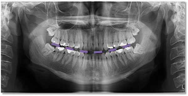
1.4 — Intraoral Imaging Is Great for Trauma, Until It Isn’t
Guideline: Intraoral radiography is useful for the evaluation of dentoalveolar trauma. If the area of interest extends beyond the dentoalveolar complex, more advanced imaging may be indicated.
What this means: Start with intraoral radiography for trauma cases — it’s effective and efficient for assessing the teeth and surrounding alveolar bone. But if clinical findings suggest the injury extends further (into the jaw, condyle, or surrounding structures), don’t hesitate to escalate to more complex imaging modalities like a panoramic radiograph or CBCT.
1.5 — Train Your Team and Prep Your Patients
Guideline: Dental staff members and operators of imaging equipment shall be trained in appropriate techniques and shall assist patients in removing objects that could affect diagnostic quality.
What this means: Quality imaging starts before the exposure. Your team needs to know how to operate every piece of imaging equipment in the office — including CBCT units. Equally important: make sure patients remove eyeglasses, jewelry, removable appliances, and anything else that could introduce artifact or compromise image quality. A retake is a second dose; prevention is better.
1.6 — Know the Risks. Explain Them.
Guideline: Clinicians, dental staff members, and operators of imaging equipment must be knowledgeable about the radiation risks associated with radiographic and CBCT imaging and be able to communicate those risks to patients.
What this means: Informed consent isn’t just a signature on a form — it’s a conversation. Your team should be comfortable discussing radiation exposure in plain language: what the doses are, how they compare to everyday background radiation, and why the imaging is clinically justified. Patients who understand the “why” are more likely to be cooperative and confident in their care.
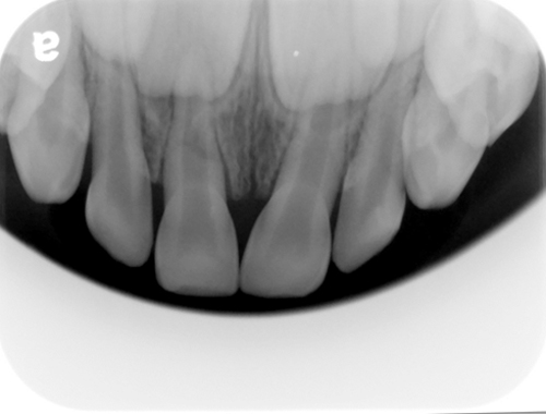
1.7 — CBCT Is Not for Caries Detection
Guideline: CBCT is not indicated for caries detection.
What this means: This one is simple and important. If you’re trying to evaluate for caries, order bitewing radiographs — not a CBCT. CBCT delivers a significantly higher radiation dose, and it does not outperform conventional radiography for detecting decay. Use the right tool for the right job.
The Bottom Line
Box 1’s general recommendations are the backbone of responsible dental imaging. They reinforce a consistent theme: imaging should be purposeful, evidence-based, and guided by clinical judgment — not habit or convenience. Understanding these guidelines isn’t just about regulatory compliance; it’s about providing better and safer care.
This is just the start of a series with more detailed information on specific indications (caries coming next) and pediatric considerations.