Since a couple of weeks ago I went over foramina on intraoral radiographs (periapical radiographs and bitewings), I thought I’d keep on going with more and this time go over canals. There is 4 again with 2 in the maxilla and 2 in the mandible; however one of the two in the mandible is possible to be seen in the maxilla.
In general (on radiographs) all canals will appear with two radiopaque lines running parallel to each other with a radiolucent center.
Nasopalatine Canal (Incisive Canal)
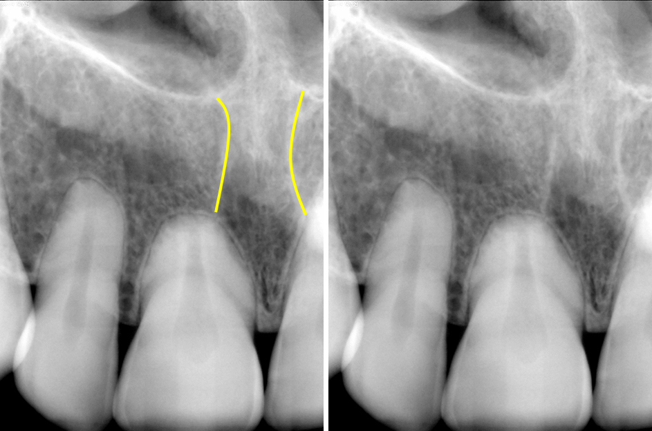
The nasopalatine canal is in the midline of the maxilla and runs from the floor of the nasal cavity to the incisive foramen which opens on the lingual between the roots of the maxillary central incisors. It is not always visible but what you want to look for is two vertical radiopaque lines. Note: sometimes only one of the borders may be seen.
Posterior Superior Alveolar Canal
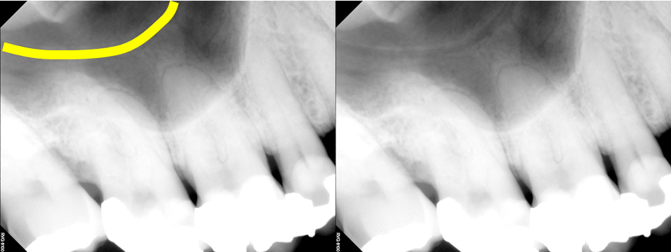
The posterior superior alveolar canal is a tricky one (and I say tricky in that this used to really make students recall their anatomy). It can be seen on posterior radiographs superimposed over the maxillary sinus. It appears as a curved horizontal radiolucent band with thin radiopaque borders in the maxillary sinus region. Note: sometimes the radiopaque borders are so thin that the radiolucent band is the more evident portion on a radiograph.
Nutrient Canals / Vascular Canals
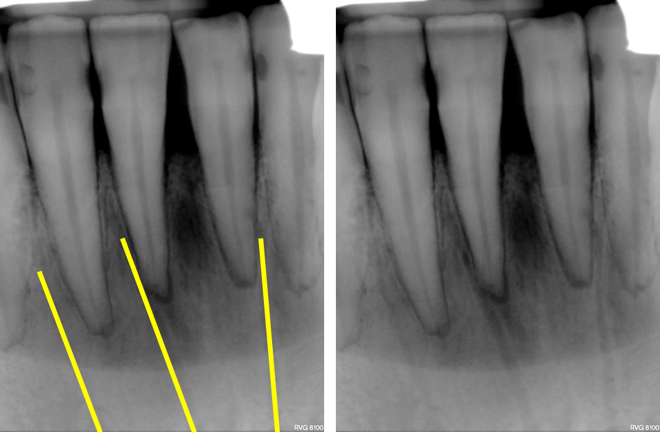
Nutrient canals are merely the accessory canals running throughout the mandible and maxilla. These canals are everywhere so theoretically they can be seen anywhere; however on 2D radiographs they are most commonly seen in the anterior mandible due to the thinness of the ridge in this location. They will appear as vertical (most common) or horizontal or really any direction radiolucent lines.
Inferior Alveolar Canal / Mandibular Canal
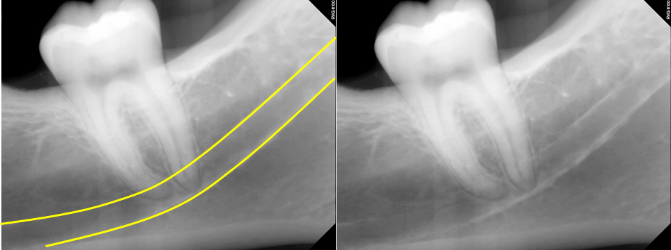
The inferior alveolar canal is probably the first canal that comes to mind when people think of canals on intraoral radiographs. This is likely due to it’s size and frequency of which it is seen. It could also be it’s such an important area in research especially regarding implant placement :). But back to what you’ll see on radiographs. It appears as two radiopaque parallel lines with a radiolucent center running oblique and horizontally inferior to or superimposed over the roots of the mandibular posterior teeth. Note: sometimes only one radiopaque line is seen. If so, it is most commonly the inferior border of the canal.
I hope this was a good refresher of canal anatomy on intraoral radiographs. I plan to keep on going with intraoral radiographs to finish off the maxilla and mandible and a few other things as well seen. If you have any comments or questions, please leave them below. Thanks and enjoy!

One thought on “Anatomy Monday: Canals on Intraoral Radiographs”
Comments are closed.