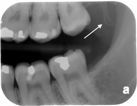
The hamulus is always a fun find when I’m working with the students on interpretations as it really forces them to recall anatomy beyond just the maxilla and mandible. The hamulus is a small bony hook that projects off the medial pterygoid plate of the sphenoid bone (that’s a tongue twister). This is typically captured on intraoral images with either excessive distal angulation or if your patient isn’t a gagger – you got the image sensor extremely far back in their mouth.
The hamulus presents as an obliquely angled radiopaque entity directly distal to the maxillary tuberosity.
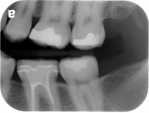
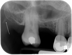
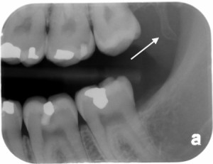
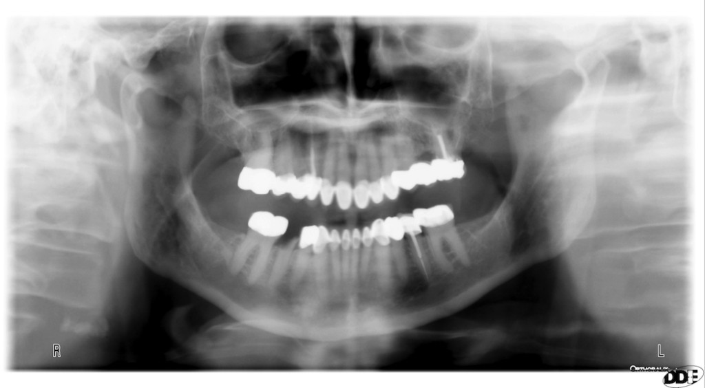
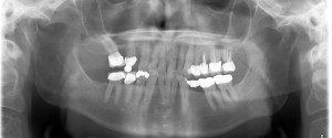
Here’s a fun example where it was visualized on a pantomograph.
If you have any questions or comments, please leave them below. Thanks and enjoy!

cool. I learned something.
🙂
is there a lateral periodontal cyst irt lower left premolars in panoramic X-ray
If you are referring to the well-defined radiolucent area between the roots of the mandibular left premolars on the pantomograph – that is the mental foramen.
Thank you so much for taking the time to create this website. It has been so helpful to my students!!
Thanks. A little behind in updating recently but pleased to hear you find it useful. 🙂