Today is the first of two posts on intraoral radiographic quality evaluation. For those who are looking for a series on pantomograph radiographic quality evaluation there are 4 posts that can be found here – ideal pantomograph quality, bad pantomograph quality – part 1, bad pantomograph quality – part 2, bad pantomograph quality – part 3.
There are 4 primary things we evaluate at the school on periapical radiographs.
1. The entire tooth or teeth to be captured are recorded on the radiograph.
This means that no portion of a tooth (crown or root) is cut off on the radiograph.
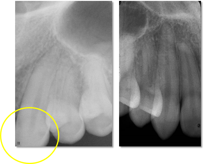
Left – circle noting the crown cut off the radiograph.
Right – entire canine is visualized on the radiograph.
2. A minimum of 2mm of bone surrounding the entire root is visible.
This includes not only at the apex of a tooth but the mesial and distal aspects as well.
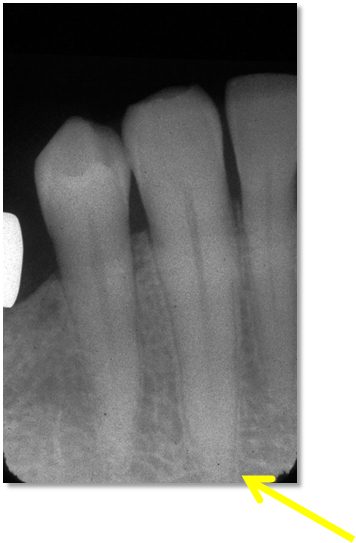
Arrow showing less than 2mm of bone around apex of the tooth.
3. Minimal overlap of the adjacent teeth.
There should be no overlap of the roots of adjacent teeth with the exception of the the canine and first premolar roots on canine periapical radiographs. Minimal overlap of adjacent crowns are acceptable as bitewing radiographs are primarily used to evaluate for interproximal decay.
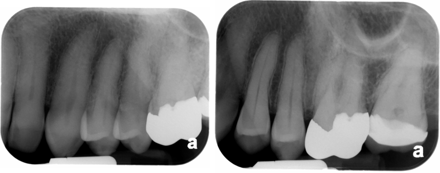
Left – note overlapping of roots and crowns causing distortion of the teeth.
Right – crowns and roots not overlapped.
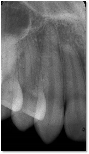
4. The roots appear longer than the crowns of the radiograph.
This means no foreshortening of the teeth on a radiograph.
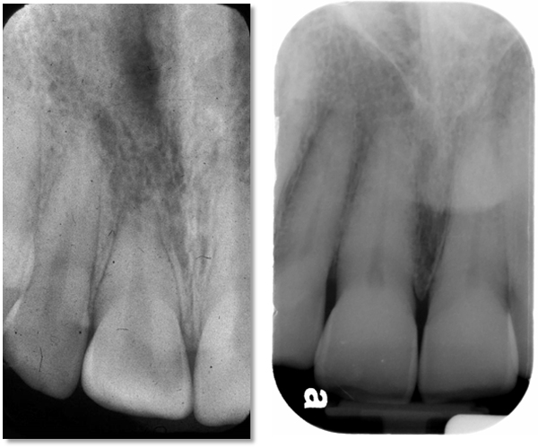
Left – note that the root and crown appear to be the same length on the radiograph.
Right – note the root appears to be twice the length of the crown which is average for this tooth.
This is a short and sweet post on the 4 main things we evaluate in the clinic. Next week will radiographic quality evaluation of bitewing radiographs. If you have any comments or questions, please leave them below. Thanks and enjoy!
