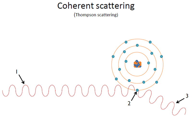
Attenuation is when the quantity of x rays is reduced by either absorption or scatter as it travels through matter. Absorption is when x ray photons ionize atoms and transfer its energy to electrons (i.e. photoelectric effect). Scattering is when the x ray photons are ejected out of the primary beam as a result of interactions with orbital electrons (i.e. coherent scattering and Compton scattering). Essentially what this is saying is that attenuation (either absorption or scatter) is how x rays are blocked/deflected and will not make it to their final destination – the image receptor.
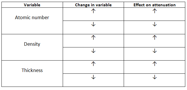
There are three primary variables that effect attenuation; atomic number, density, and thickness. The higher the atomic number of an object, the more x rays it will stop. The higher density an object has, the more x rays it will stop. And lastly, the thicker an object is, the more x rays it will stop from reaching the image receptor.
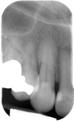
For example, let’s compare enamel versus air. Enamel is thicker, has a higher density and higher atomic number than air. These all lead to enamel stopping more x rays from making it to the image receptor versus air. On the radiograph below, you can see how the final image of enamel compares to air. For more practice on this topic, choose two different structures (i.e. metal and enamel, bone and the root canal space, etc.) and compare them. Use the radiograph below to see how attenuation effects the final image. 🙂
This is a short post on attenuation basics and how it plays a role in radiography. The three specific methods of attenuation (Compton scatter, Photoelectric effect and Coherent scatter) will be covered in the upcoming weeks. If you have any questions, please let me know. Thanks and enjoy!
One thought on “All about attenuation of x rays”
Comments are closed.