This is part 2 (posterior) of anatomy on intraoral radiographs.
Mandible
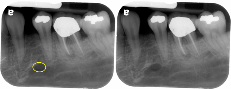
The mental foramen appears as a round to oval radiolucent area near the apex of the second premolar.
The inferior alveolar nerve canal (mandibular canal) appears as radiolucent band with two thin radiopaque lines running parallel to each other (superior and inferior). If only one border it visible, it is more likely the inferior border.
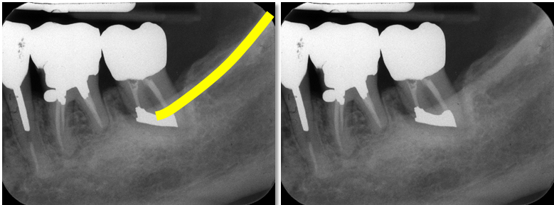
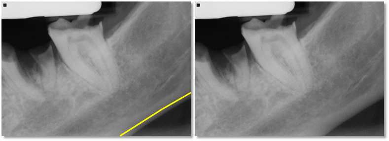
The external oblique ridge (external oblique line) appears a thick radiopaque line that runs obliquely as it descends and superimposes over the roots of the molars.
The mylohyoid ridge appears as a thick radiopaque line frequently seen near the roots/apices of the posterior teeth.
The submandibular salivary gland fossa appears as an area of more radiolucent bone inferior to the mylohyoid ridge.
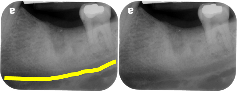
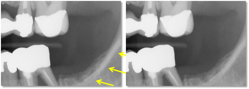
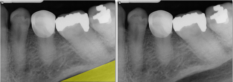
The inferior border of the mandible appears as a thick radiopaque band.
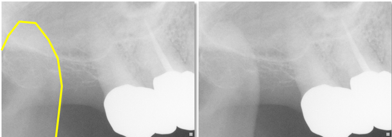
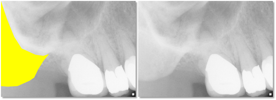
The coronoid process is seen on maxillary molar periapicals. It appears as a triangular radiopaque area superimposed over the maxillary molars and tuberosity.
Maxilla
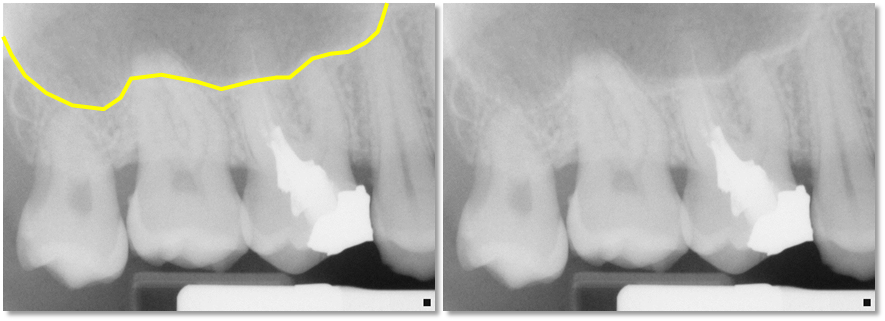
The border of the maxillary sinus appears as a thin radiopaque line superior/superimposed over the roots of the posterior teeth. The maxillary sinus appears as a radiolucent area superior to the border of the maxillary sinus.
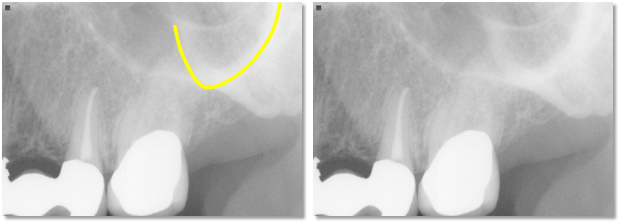
The zygomatic process of the maxilla appears as a U, V or J shaped radiopaque line. It is superior to the first and second molars.
The zygomatic bone appears as a radiopaque area distal to the zygomatic process of the maxilla.
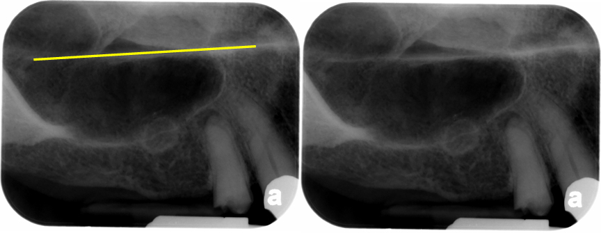
The floor of the nasal cavity appears as a thin straight radiopaque line superimposed over the maxillary sinus.
I hope you find this second post on intraoral anatomy informative. While I didn’t cover all the anatomy visible, these are the most commonly seen entities. If you have any questions, please let me know.
Coming next (week of July 9 – 13th) Anatomy on Pantomographs Part 1.
Enjoy!

Awesome
very good images and explanations, I’m studying for my national dental boards and this is very helpful
Thanks and I’m glad you found this useful. 🙂
Thank you very much !
Very informative ,I am a DH student in my first semester. Very thank your post. It is very helpful for my radiology exam.
Thank you. 🙂