1 min read 8

Lateral Cephalometric Skull Anatomy – Part II

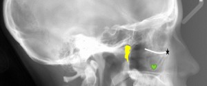
Onto part 2 of lateral cephalometric skull anatomy frequently used in orthodontics. Again identified on a dry skull first followed by the radiograph. Orbitale (black star) – The lowest point of the orbital margin (also the anterior point for the Frankfort horizontal plane). Key ridge (green heart) – The inferior…
1 min read 7

Anatomy Monday: Anterior Nasal Spine (maxilla)

This week’s anatomy is the anterior nasal spine which is found in the midline at the floor of the nasal cavity as a bony protuberance where the right and left maxilla meet (as seen on the skull below – orange arrow).On radiographs, this will appear as an inverted triangle or…
1 min read 6

Case of the week: Talon cusp with clinical photo

This week I have a case of a large talon cusp on a maxillary central incisor along with a clinical photo.  A talon cusp is a hyperplasia of the cingulum of a tooth and typically classified as a variant of normal anatomy.  In the anterior, it will present as a…
1 min read 0

Anatomy Monday: Coronoid Process (mandible)

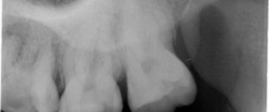
The coronoid process is the superior aspect of the anterior portion of the ramus of the mandible.  It is visible on intraoral radiographs but not mandibular periapical radiographs instead it is seen on maxillary molar periapical radiographs.  This typically due to the posterior placement of the image receptor and holder…
1 min read 0

Case of the week: External resorption of an impacted tooth

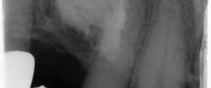
This week is a neat case of an impacted maxillary canine with external resorption occurring on the crown.  When looking at these radiographs note that there is no longer an identifiable follicle around the crown of the tooth.  Also note that the areas where there is tooth loss of the…