1 min read 0

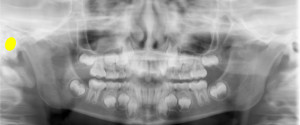
Anatomy Monday: External Auditory Meatus

This week is the external auditory meatus.  The external auditory meatus is the external opening to the auditory canal. It presents as a well-defined round to ovoid radiolucent area lateral to the condyle. If you have any questions or comments, please leave them below. Thanks and enjoy!
1 min read 3

Case of the Week: Antrolith

This week is a case of calcifications in the maxillary sinus commonly referred to as antroliths.  This case is a CBCT case seen as multiple well-defined radiopaque entities near the floor of the right maxillary sinus. If you have any questions or comment, please leave them below. Thanks and enjoy!
1 min read 0

Case of the Week: Mucous Retention Pseudocyst

This week I have a case of a mucous retention pseudocyst on a pantomograph. Mucous retention pseudocysts are incidental findings that do not require treatment. They are most commonly found in the maxillary sinuses followed by the sphenoid sinuses. Note the rounded radiopaque dome in the right maxillary sinus. If…
1 min read 0

Case of the Week: Oligodontia

This week I have a case of developmentally missing teeth (oligodontia) along with the last educational video in my library (to date). 🙂 This case shows all four second premolars did not develop with retained primary second molars. And for the last of this years educational videos on oligodontia by…
1 min read 0

Case of the Week: Pulp Stones

This week is a case of two elongated pulp stones along with an educational video. The case shows an oblong radiopaque mass in the pulp chamber and root canal space of the canine and first premolar. Note how the chamber and canal space is enlarged around the radiopaque mass. Here’s…