1 min read 0

Case of the Week: Palatal Tori

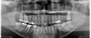
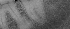
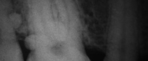
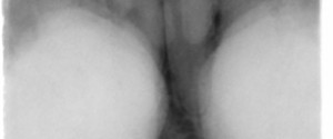
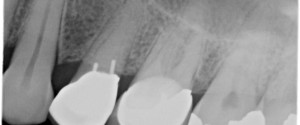
This week I have a case of palatal tori evident not only on a periapical radiograph but a pantomograph as well.  On the periapical radiograph it appears as a radiopaque mass superior to the maxillary teeth. The pantomograph shows a well-defined horizontal radiopaque band directly inferior to the floor of…
1 min read 0

Case of the Week: Talon Cusp

This week I have a case of a talon cusp so large it’s evident on a pantomograph.  While there is more than one talon cusp evident on this radiograph, it’s the one on the maxillary left lateral incisor that is nearly as big as the crown that’s amazing :).  Note…
1 min read 0

Case of the Week: Hypercementosis

This week is a fun case of hypercementosis at the apex of a molar root.  Note on the distal root of the first molar a radiopaque mass with a thin radiolucent line around it. This shows that the mass is attached to the root surface with the periodontal ligament space…
1 min read 0

Case of the Week: Enamel Pearls

I have to send a big thank you out to Dr. Marc I Semper for sharing this case of enamel pearls with me.  He was also kind enough to share a link to a great overview page about enamel pearls by Marco A Versiani.  Below I’ll let you find the…
1 min read 0

Case of the Week: Mandibular Tori

This week is a more common radiographic finding; mandibular tori. This case is bilateral and dense mandibular tori.  It’s pretty obvious so I’m just going to let you take a look for yourself. 🙂 And here’s another educational video from Dental Class of 2015 students Paul Favela and Ben Winston.…