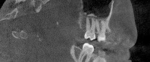
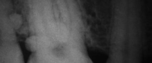
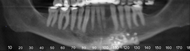
This week is a case of calcifications in the maxillary sinus commonly referred to as antroliths. This case is a CBCT case seen as multiple well-defined radiopaque entities near the floor of the right maxillary sinus.
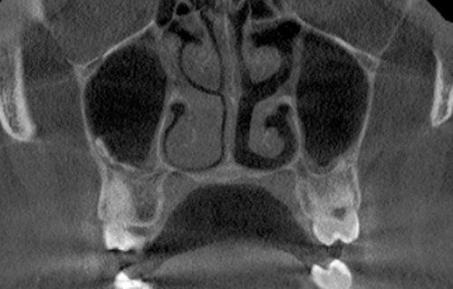
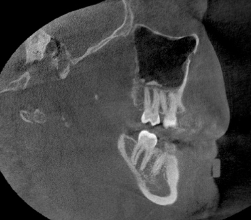
CBCT coronal view – multiple radiopaque entities near the floor of the right maxillary sinus and a thin layer of increased mucosal thickening of both the right and left maxillary sinusesCBCT sagittal view – multiple radiopaque entities near the floor of the maxillary sinus and mucosal thickening
If you have any questions or comment, please leave them below. Thanks and enjoy!
3 thoughts on “Case of the Week: Antrolith”
Nice slices, Shawneen, as this is a very rare occurence. Cheers, Marc
Nice images, How about patient’s right nasal cavity (nares)! it is almost blocked! Referral to ENT?
The soft tissue of the conchae are enlarged with very little space for air to move through that side of the nasal cavity. A referral to an ENT would be recommended especially if the patient is having airway or breathing issues.