1 min read 0

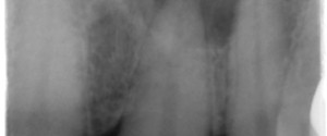
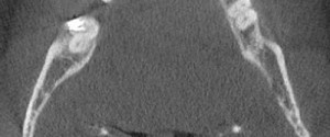
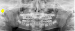
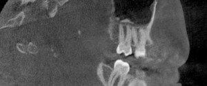
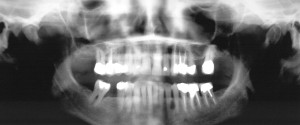
Case of the Week: Lateral Periodontal Cyst

This week is a case of a cyst most commonly found as an incidental finding on radiographs; the lateral periodontal cyst. It is most commonly found in the mandibular canine/premolar region. This case shows a well-defined ovoid radiolucent entity between the mandibular left canine and first premolar roots. If you…
1 min read 2

Case of the Week: Radicular / Periapical cyst

Now before I get to the actual case I want to make sure that you realize that a radicular/periapical cyst is diagnosed with a histopathological slide and not by radiographs alone.  There are certain features of a radicular/periapical cyst that may cause you to put it higher on a differential…
1 min read 2

Case of the Week: Tonsiliths

This week is another CBCT case of calcifications, this time in the tonsils.  On CBCT images, these are seen on the axial views lateral to the airway and on coronal views medial to the ramus of the mandible.  They are well-defined irregular shaped radiopaque entities. They may be single or…
1 min read 0

Anatomy Monday: External Auditory Meatus

This week is the external auditory meatus.  The external auditory meatus is the external opening to the auditory canal. It presents as a well-defined round to ovoid radiolucent area lateral to the condyle. If you have any questions or comments, please leave them below. Thanks and enjoy!
1 min read 3

Case of the Week: Antrolith

This week is a case of calcifications in the maxillary sinus commonly referred to as antroliths.  This case is a CBCT case seen as multiple well-defined radiopaque entities near the floor of the right maxillary sinus. If you have any questions or comment, please leave them below. Thanks and enjoy!