1 min read 0

Case of the week: Calculus with clinical photo

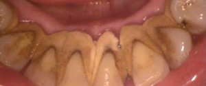
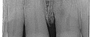
This week I have a case of some serious calculus on the mandibular anterior teeth.  I have both a radiograph and a clinical photo as well.  Calculus appears a radiopaque ‘growths’ on the mesial and/or distal surfaces of teeth.  It is typically found near the cemento-enamel junction but can occur…
1 min read 0

Case of the week: Multirooted Maxillary Central Incisor

This weeks case was a really cool find.  All the endo residents wanted to treat it as it would be an amazing boards case.  Granted the tooth is in good health and no endodontic therapy is needed.  So here is a multirooted maxillary central incisor. Note the two orange arrows…
1 min read 3

Case of the week: Fusion

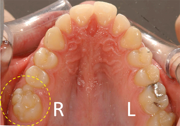
This week is a neat case of fusion with some clinical photos as well.  Fusion is when two adjacent developing teeth fuse into one large tooth.  This results in an overall tooth count of 15 for an arch.  Gemination, which can have a similar appearance, results in an overall tooth…
1 min read 4

Case of the week: Double Lingual Foramina

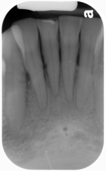
This weeks case is an interesting variant of normal anatomy – two lingual foramina seen on a periapical radiograph.  Some patients can have up to three lingual foramina. Note the two well-defined, circular radiolucent entities inferior to the mandibular central incisors.  The superior one is larger than the inferior one.…
1 min read 4

Case of the week: Chronic Apical Periodontitis

This week is a follow up to last week with a case of chronic apical periodontitis.  The primary difference of chronic apical periodontitis versus acute apical periodontitis is the presence of sclerotic bone formation.  This sclerotic bone formation (sometimes referred to as sclerosing osteitis) may present in one of three…