5 min read 0

Anatomy Monday: Anatomy on Mandibular Periapical Radiographs

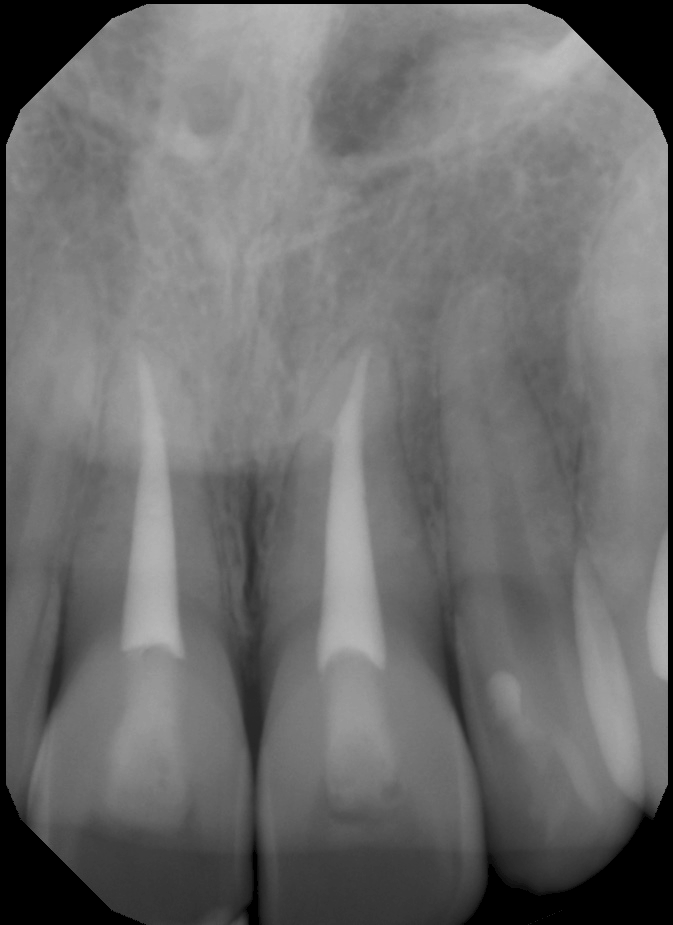
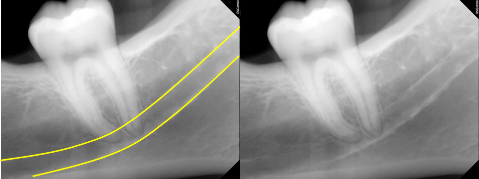
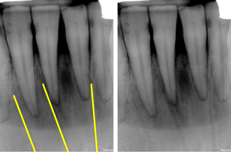
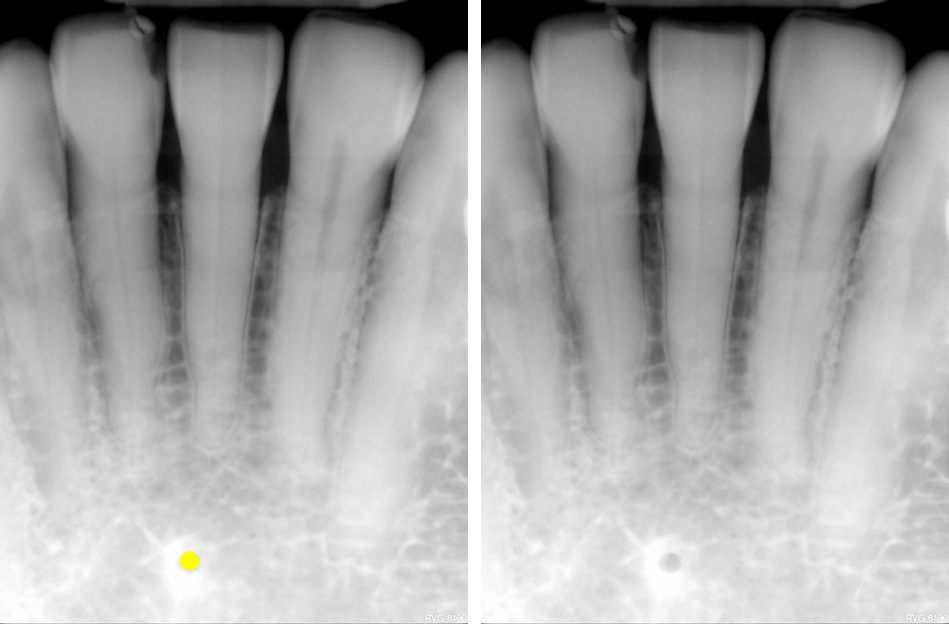
It’s been a while since I’ve updated anatomy seen on intraoral radiographs so I’m continuing with all the anatomy visible on mandibular periapical radiographs. Some of these were seen in the recent posts on canals and foramina but this will be the one stop spot for everything mandibular periapical radiographs.…
3 min read 1

Anatomy Monday: Canals on Intraoral Radiographs

Since a couple of weeks ago I went over foramina on intraoral radiographs (periapical radiographs and bitewings), I thought I’d keep on going with more and this time go over canals. There is 4 again with 2 in the maxilla and 2 in the mandible; however one of the two…
3 min read 1

Anatomy Monday: Foramina on Intraoral Radiographs

Today I thought I’d go with a little refresher of foramina visible on intraoral radiographs specifically periapical radiographs.  There will be a total of 4 I’m covering, 2 each in the maxilla and mandible.  So onto the maxilla first. Incisive Foramen The incisive foramen is the inferior opening of the…
1 min read 0

Anatomy Monday: Zygomatico-temporal suture

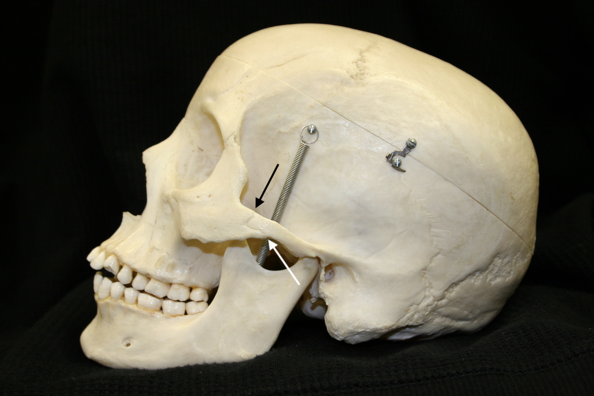
I thought I’d have some fun with today’s anatomy by posting one that is not always visualized on pantomographs and sometimes is mistaken for a fracture when it is visualized.  The zygomatico-temporal suture is the junction of the zygomatic bone and temporal bone in the zygomatic arch.  It varies on…
1 min read 0

Anatomy Monday: Cervical Vertebra 1 (C1 or Atlas)

Since I showed a case of a posterior cleft of C1 last week I thought I’d follow up with some key anatomy of C1 as seen on cone beam computed tomography (CBCT) images. These are all axial views that have been rotated as such to show the entire ring on…