Now the answer from Tuesday.
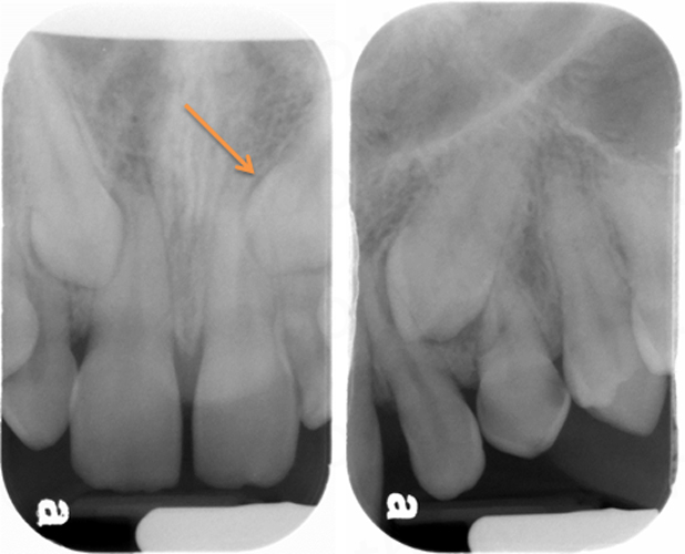
Where is the accessory root (white arrow) in relation to the distal root (yellow arrow) of the first molar?
Before starting to use the image shift principle it is important to know/remember two key points
- Images move in the opposite direction from the movement of the source.
- Images of objects farther from the image receptor (sensor, plate, film) will move more (aka objects (images) more facial/buccal will appear to move more).
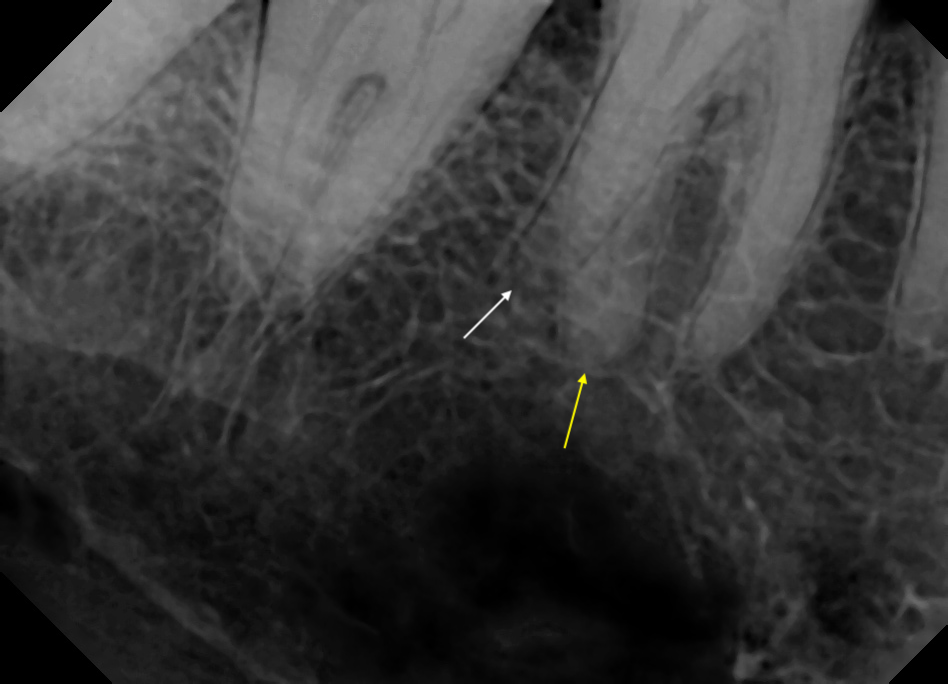
On these radiographs, we are going to use the distal root (yellow arrow) of the first molar as our stationary object as it is seen on both radiographs and appears to move in comparison to the accessory root.
The next step is to determine what angle change is obvious between the two radiographs? Positive vertical angle, negative vertical angle or horizontal angle.
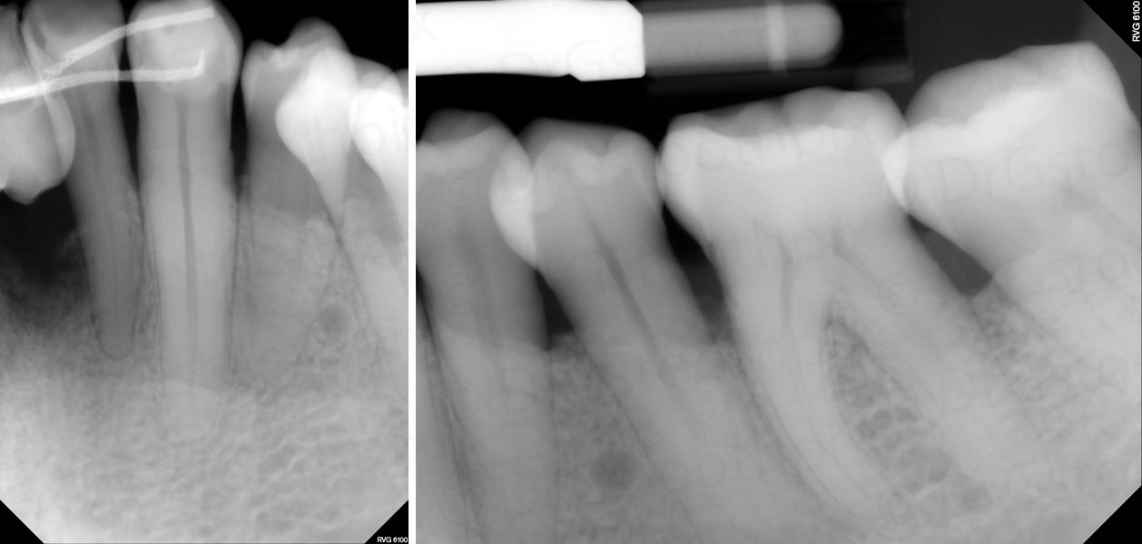
The most obvious angle change is the horizontal angle between the two radiographs. Starting with the mandibular right molar periapical radiograph and moving to the mandibular right molar/premolar periapical radiograph, the horizontal angle decreases meaning the source of radiation (tubehead) moves anteriorly. According to point 1 above, this means the images move posterior.
Looking at the second radiograph (mandibular right molar/premolar periapical radiograph), we need to compare the image movement of the accessory root (white arrow) versus the distal root (yellow arrow) to see which object moved more posterior following point 2 listed above.
The accessory root (white arrow) appears to be more posterior on the mandibular right molar/premolar periapical radiograph meaning it is farther from the image receptor compared to the distal root (yellow arrow).
This gives us an answer of the accessory root (white arrow) being to the facial of the distal root (yellow arrow) of the first molar.
SLOB (Same-Lingual, Opposite-Buccal)
We will use the same objects as above (unknown object = accessory root and fixed object = distal root).
Next, we need to determine which direction we are moving from the mandibular right molar periapical radiograph to the mandibular right molar/premolar periapical radiograph and the answer would be – mesial.
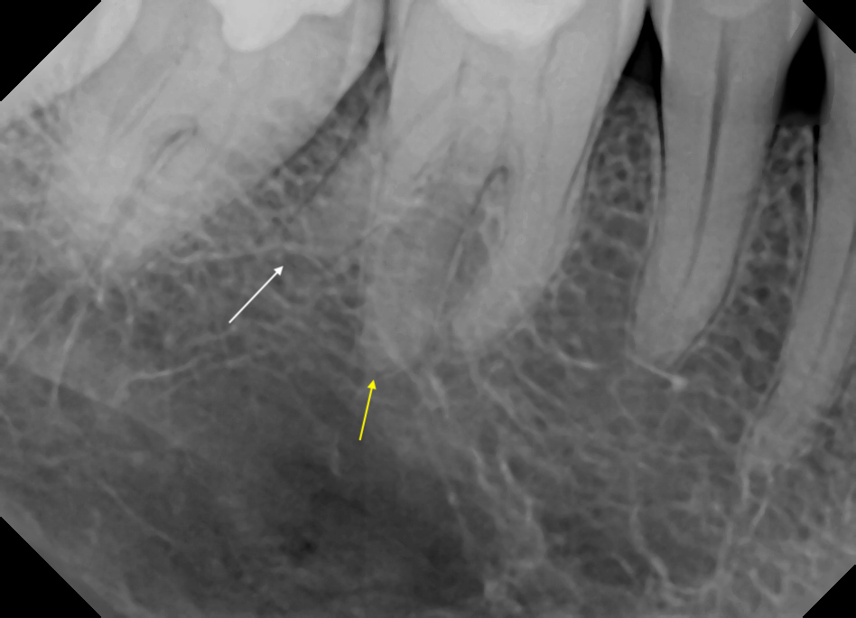
On the mandibular right molar/premolar periapical radiograph determine what direction does the accessory root appear to have moved in relation to the distal root of the first molar – distal.
Here is where the acronym comes into play. Did the unknown object (accessory root) move in the SAME direction as the radiographs or in the OPPOSITE direction?
Our answer is – opposite and the acronym states that opposite is buccal, so the accessory root is to the buccal of the distal root of the first molar.
If you have any questions or comments, please leave them below. Thanks and enjoy!