This how was that missed post is showing a case of a mandibular fracture missed on a 2D image but easily seen on a CT scan.
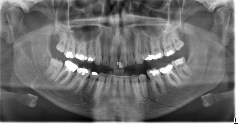
Initial interpretation: Obvious radiolucent line extending from mandibular left premolar region to the inferior border of the mandible. There is a discontinuity of the inferior border at this spot. The radiograph shows a single mandibular fracture of the left body.
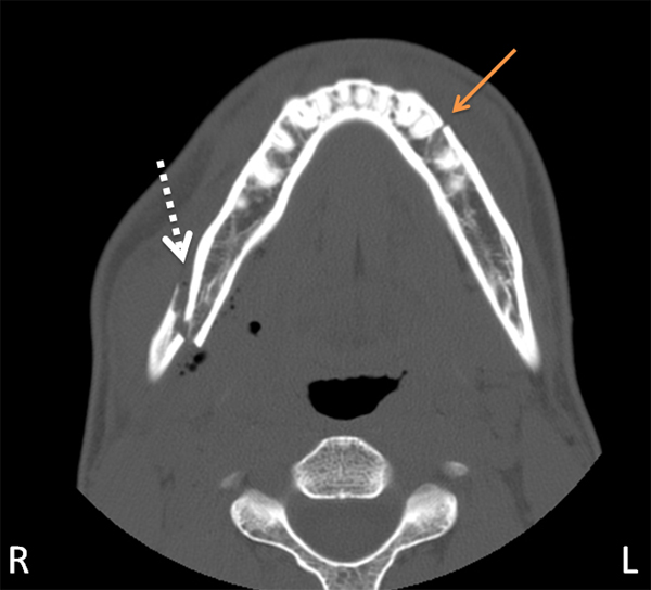 Axial view showing two fractures of the mandible (white dotted and orange arrows)
Final Interpretation: (after reviewing CT scan) There is not just one but two fractures of the mandible creating a segmental fracture of the mandible. While the left fracture (orange arrow above) was easily seen on the pantomograph, the right fracture (white dotted arrow) was not as obvious.
Lesson learned: The second fracture (right) shows the least common way fractures present on radiographs – increased radiopacity due to overlapping of the two segments. This case was good in reminding me of that specific radiographic finding of fractures. After seeing the CT scan and going back to look at the pantomograph, you will note there is a radiopaque U shaped entity near the right antegonial notch region. This is where the two segments of the mandible overlapped. Go here for a reminder of the 4 radiographic findings of fractures.
If you have any questions about this case or radiographic findings of fractures, please let me know. Thanks and enjoy!

Auch dieses blonde Teenie macht da keine Ausnahme.