This week has a warning…
Be forewarned that there are some images that are not suitable for viewers of all ages (it gets a little bloody).
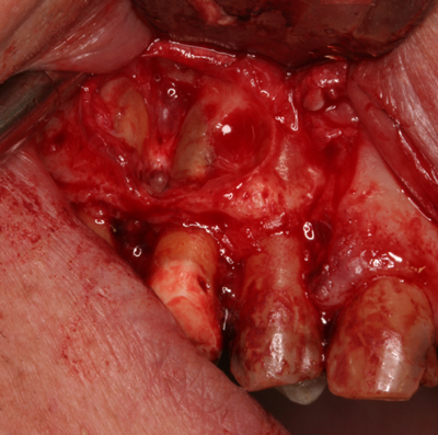
Ok, now that I’ve said that I’ll continue on with this amazing case of a keratocystic odontogenic tumor (which used be called an odontogenic keratocyst – name change due to tumor-like growth instead of cystic growth). This case was present in the posterior maxilla and I have some clinical photos of the removal showing the actual lesion. This location is not the most common location as keratocystic odontogenic tumors occur more frequently in the posterior mandible. This entity contains keratin which has a yellow cheesy appearance and thsis yellowish color is evident on the clinical picture below. Whenever considering a keratocystic odontogenic tumor on a radiograph, it should be noted that the following entities should also be included on a differential list.
2. Odontogenic myxoma
3. Central hemangioma
Now onto the case.
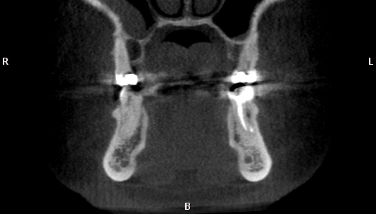
Coronal CBCT slice showing well-defined radiolucent entity in the right posterior maxilla palatal to the tooth.
Clinical photos – left image shows yellowish coloring of neoplasm – right image shows exposed roots after removal of the neoplasm.
Enjoy (and I hope the pictures weren’t too disturbing for you)!


It is absolutely necessary CBCT for the diagnostic or could be seen in a pano or ceph?
CBCT is helpful to give the practitioner more information and aid in surgery for removal or biopsy. Not necessary but many like to have for these reasons.
Other 2D radiographs (such as pantomographs and cephalometric skulls) also provide additional information about the area in question.
Dear Dr.
It can be seen in the CTCB that the tumour is in the palatal side, but in the clinical pictures, it is from the buccal side, so is there any mistake here.
The lesion on the buccal side was the initial lesion. The lesion on the CBCT on the palatal side was a secondary lesion that was found on the scan after the initial surgery when evaluating to make sure it was completely removed.