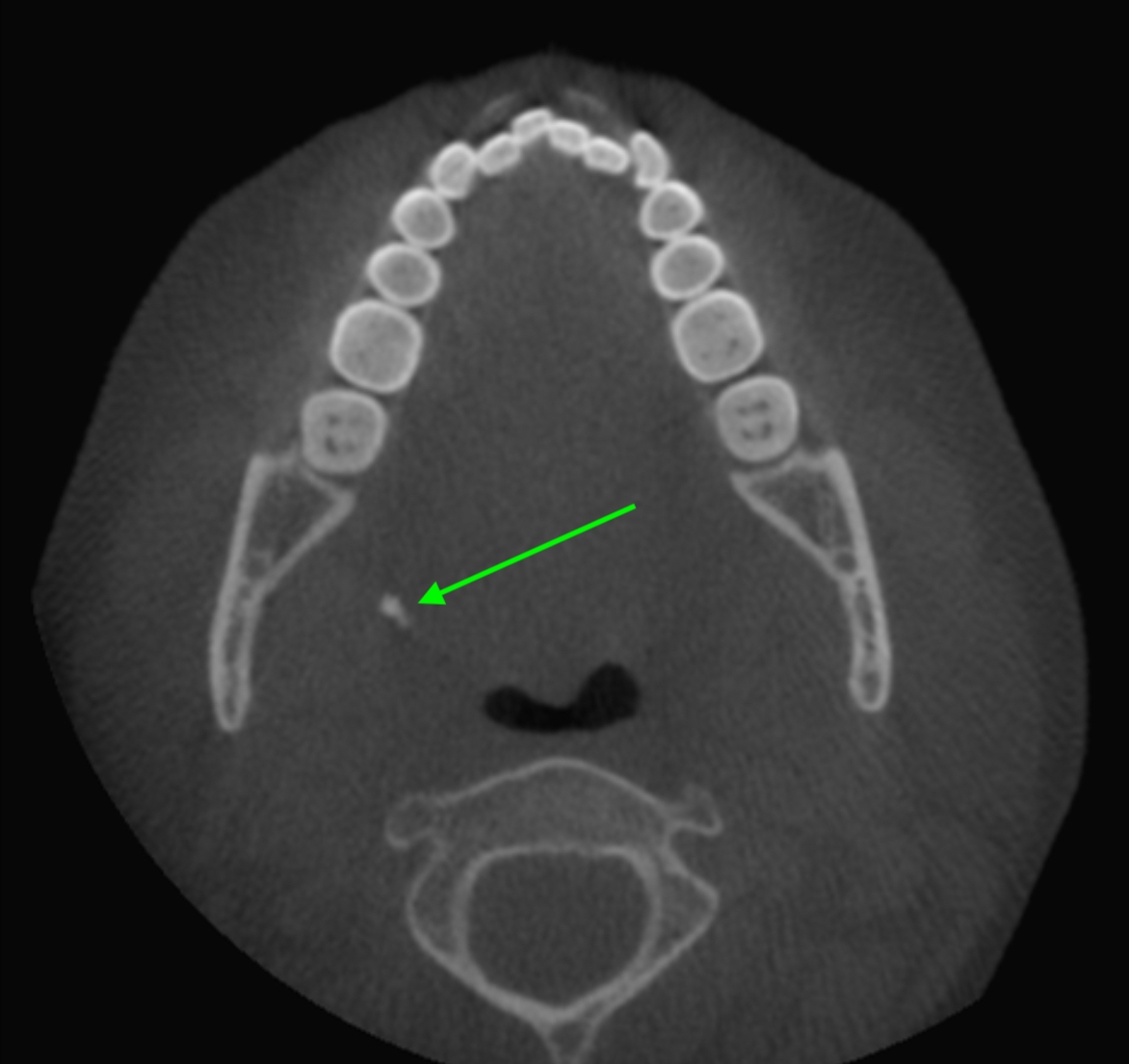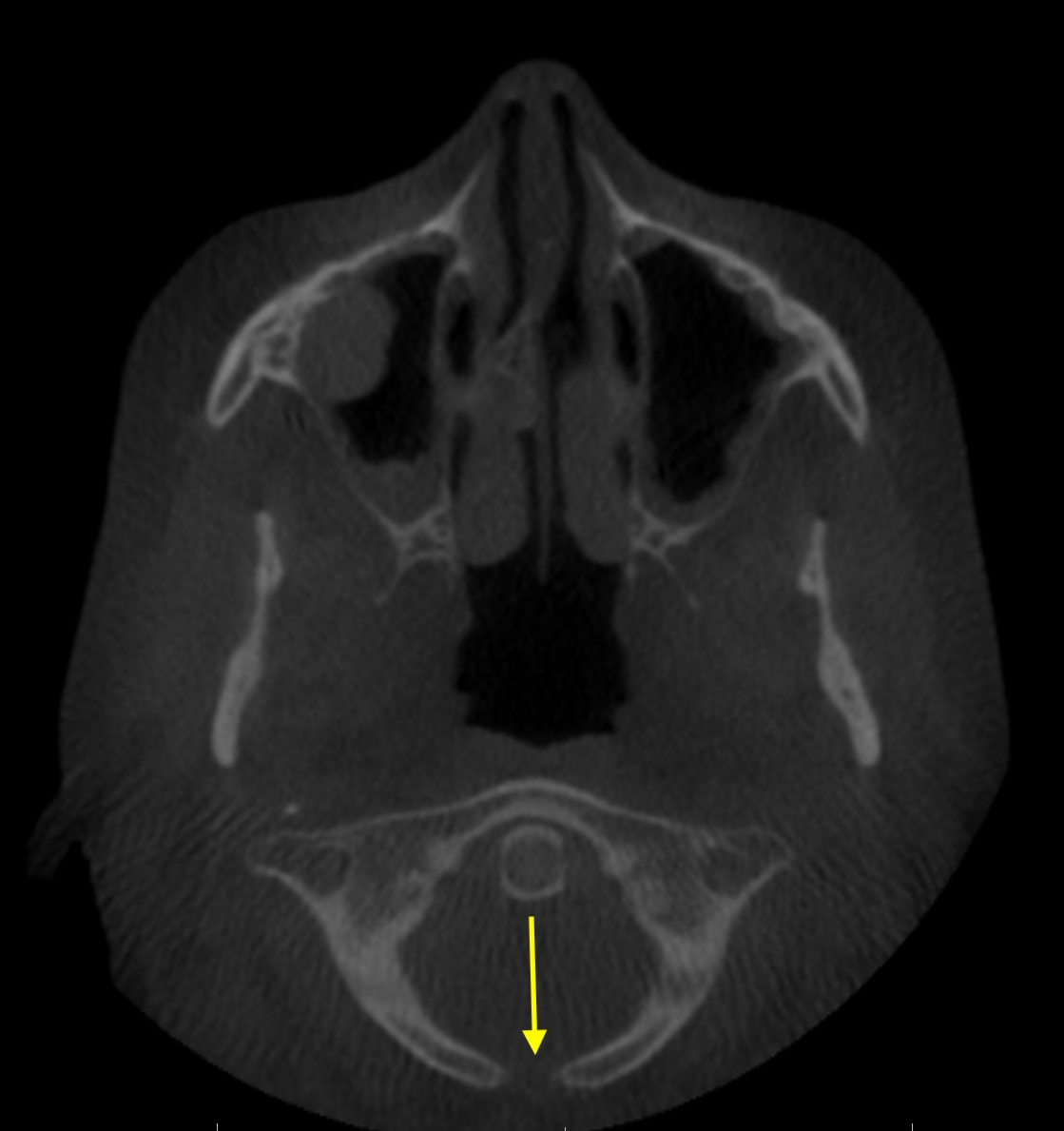
This week I have a developmental anomaly where the posterior arch of C1 does not completely fuse together. This can be seen best on axial views on cone beam computed tomography (CBCT). Due to the angle of the scan you won’t likely see the entire arch as I have shown here unless you rotate the scan to see it all together.

For more information on other anomalies of C1 (there’s an anterior cleft as well which I’ll add in the future) check out Radiopaedia’s post.
Thanks and enjoy!