Skip to content
Dr. G's Toothpix

Dr. G's Toothpix

An oral radiology source for dental professionals.

  • START HERE
  • Radiology Reporting Service
    • Services / Pricing
    • How To Upload A Case
    • Sample Report
    • Contact
  • Describing Radiographic Lesions
  • Radiographic Interpretation
    • Benign Neoplasms
      • Ameloblastoma
      • Cementoblastoma
      • Compound Odontoma
      • Enostosis
      • Focal Idiopathic Osteosclerosis
      • Odontogenic Keratocyst
    • Cysts of the Jaws
      • Buccal Bifurcation Cyst
      • Dentigerous Cyst
      • Lateral Periodontal Cyst
      • Nasopalatine Canal Cyst
      • Periapical / Radicular cyst
      • Simple Bone Cyst
    • Maxillary Sinuses
      • Antral Exostoses / Antral Projection
      • Antrolith
      • Mucositis
      • Mucous Retention Pseudocyst
      • Sinusitis
    • Periapical Inflammatory Diseases
      • Rarefying Osteitis
      • Sclerosing Osteitis
      • Widened Periodontal Ligament Space
    • Skeletal Developmental Abnormalities
      • Cleft Palate
      • Stafne Defect
    • Soft Tissue Calcifications / Ossifications
      • Antrolith
      • Arterial calcifications
      • Ossified Stylohyoid Ligament
      • Osteoma Cutis
      • Rhinolith
      • Sialolith
      • Tonsiliths
    • Tooth anomalies
      • Abrasion
      • Amelogenesis Imperfecta
      • Ankylosis
      • Attrition
      • Dens Evaginatus
      • Dens Invaginatus
      • Dentinogenesis Imperfecta
      • Ectopic Eruption
      • Enamel Pearl
      • External Resorption
      • Gemination
      • Hypercementosis
      • Internal Resorption
      • Macrodont
      • Microdont
      • Oligodontia
      • Pulp Stone
      • Pulpal Sclerosis
      • Supernumerary teeth
      • Talon cusp
      • Taurodontism
      • Transposition
    • Trauma to Teeth and Jaws
      • Coronal fractures
      • Fibrous Scar
      • Mandibular Fractures
      • Root fractures – Horizontal
      • Root fractures – Vertical
  • About
    • Disclaimer
    • Publications

Locate the Object: February 2015 – 17

Posted on February 26, 2015February 24, 2015 by Dr. Shawneen Gonzalez

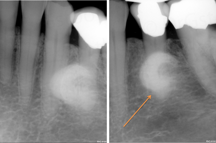
image shift 4-1

Where is the radiopaque mass (orange arrow) in relation to the mandibular left second premolar (#20)?

Answers coming later today. If you have any questions or comments, please leave them below. Thanks and enjoy!
Posted in Radiographic InterpretationTagged image shift, locate the object, SLOB rule

Post navigation

Previous: Locate the Object: February 2015 – 16
Next: Locate the Object: February 2015 – 16 ANSWER

Looking for something?

Post topics

anatomy anterior nasal spine artifacts caries caries interpretation CBCT CBCT anatomy CBCT interpretation common radiographic errors cysts of the jaws digital radiography external resorption floor of the nasal cavity image shift impacted tooth intraoral radiograph anatomy intraoral radiographic technique lateral cephalometric skull anatomy LESION lingual foramen locate the object maxillary sinuses mental foramen microdont oral radiology websites pantomograph anatomy pantomograph positioning phosphor plates pulpal/apical findings radiation physics radicular / periapical cyst radiographic interpretation radiographic quality evaluation radiographic technique rarefying osteitis resorption sensors SLOB rule soft tissue calcifications / ossifications supernumerary teeth supernumerary tooth tooth anomalies trauma to teeth and jaws videos x ray

Looking for something?

Recent Posts

  • Imaging for Periodontal Disease: What the 2026 ADA & AAOMR Guidelines Say
  • Imaging for Caries Detection: What the 2026 ADA & AAOMR Guidelines Say
  • Understanding the 2026 ADA & AAOMR General Recommendations for Dental Radiography and CBCT
  • Radiographic Interpretation: ‘hydraulic’ appearance
  • Updated Page: Hypercementosis

Post topics

anatomy anterior nasal spine artifacts caries caries interpretation CBCT CBCT anatomy CBCT interpretation common radiographic errors cysts of the jaws digital radiography external resorption floor of the nasal cavity image shift impacted tooth intraoral radiograph anatomy intraoral radiographic technique lateral cephalometric skull anatomy LESION lingual foramen locate the object maxillary sinuses mental foramen microdont oral radiology websites pantomograph anatomy pantomograph positioning phosphor plates pulpal/apical findings radiation physics radicular / periapical cyst radiographic interpretation radiographic quality evaluation radiographic technique rarefying osteitis resorption sensors SLOB rule soft tissue calcifications / ossifications supernumerary teeth supernumerary tooth tooth anomalies trauma to teeth and jaws videos x ray

Recent Posts

  • Imaging for Periodontal Disease: What the 2026 ADA & AAOMR Guidelines Say
  • Imaging for Caries Detection: What the 2026 ADA & AAOMR Guidelines Say
  • Understanding the 2026 ADA & AAOMR General Recommendations for Dental Radiography and CBCT
  • Radiographic Interpretation: ‘hydraulic’ appearance
  • Updated Page: Hypercementosis

Related Posts

  • Radiographic Interpretation

Anatomy on Radiographs: Pantomographs Part 2

  • Dr. Shawneen Gonzalez
  • July 19, 2012
  • 2 min read
  • 10
  • Radiographic Interpretation

Locate the Object: July 2015

  • Dr. Shawneen Gonzalez
  • July 29, 2015
  • 1 min read
  • 4
  • Radiographic Interpretation

Educational Video: Scleroderma

  • Dr. Shawneen Gonzalez
  • July 31, 2014
  • 1 min read
  • 0
  • Radiographic Interpretation

Educational Video: Sinusitis

  • Dr. Shawneen Gonzalez
  • August 14, 2013
  • 1 min read
  • 0

(c) 2010 – 2014 DrGstoothpix

This site is an educational resource of radiographic findings in dentistry. This information is meant as a learning tool and not a diagnostic tool. Any and all diagnoses should be made by your personal physician or dentist after reviewing clinical and radiographic findings and medical and dental health history. The views expressed in this blog/website are my own and do not reflect the views of UNMC.

(c) DrGstoothpix 2010 – 2020

This site is an educational resource about oral and maxillofacial radiology. This information is meant as a learning tool and not a diagnostic tool. Any and all diagnoses should be made by your personal physician or dentist after reviewing clinical and radiographic findings and medical and dental health history. The views expressed in this blog/website are my own and do not reflect the views of my employer.

Copyright © 2026 Dr. G's Toothpix Theme: Flawless Blog By Adore Themes.