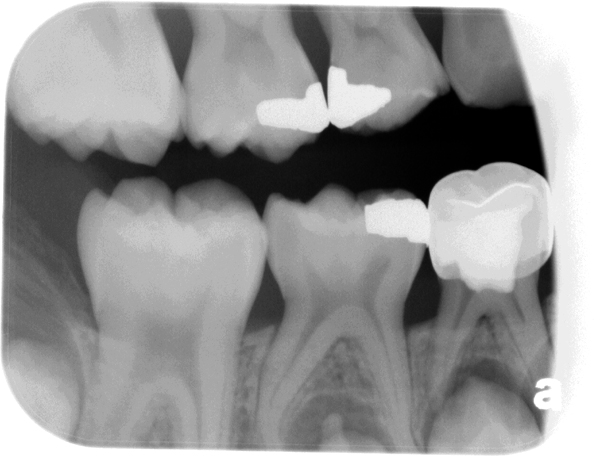
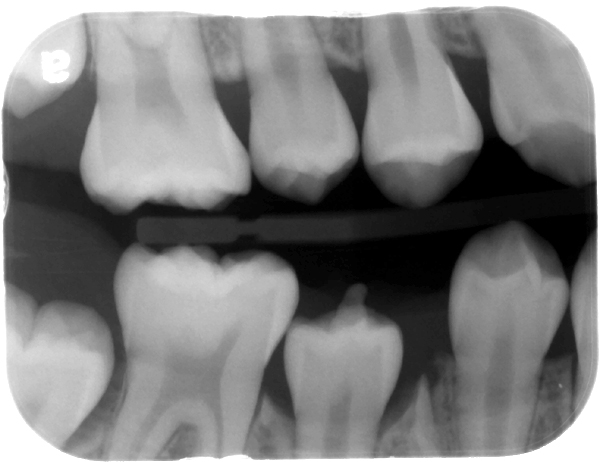
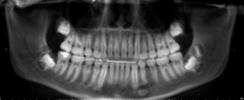
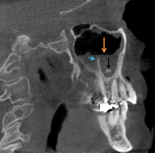
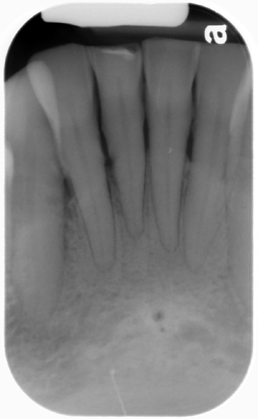
This week is a really cool case of a dens evaginatus erupting onto the plane of occlusion. Dens evaginatus is an outfolding of the enamel from the occlusal surface of a tooth, which is opposite of dens invaginatus. On this case, look at the mandibular second premolar which appears to have a vertical projection of enamel in the follicle.
The second radiograph is after the exfoliation of the primary teeth with more of the crown of the second premolar evident.
If you have any questions or comments about this case, please leave them below. Thanks and enjoy!
Hi Dr. G, I’m a dental student at the University of Toronto in Canada and find your website to be really helpful as a supplement to our lecture notes. Thanks so much for making this site! Just a question on the first radiographic image. Why is the second primary molar more radioopaque than the rest of the teeth, especially where the pulp chamber is? Also, do we tend to expect the permanent tooth that’s replacing the primary to also be affected? Thanks so much in advance!
Thanks and I’m glad you are finding the site informative. 🙂
Are you asking about the radiopaque filling material on the mandibular first primary molar? If so, that’s endodontic material used for primary teeth which is typically only applied in the pulp chamber. For the permanent tooth replacing the first primary tooth if an inflammation is present in the bone near the developing premolar it can result in altered morphology of the crown of the permanent tooth which is referred to as Turner’s hypoplasia.
Please let me know if this is what you were asking and tell Dr. Ernest Lam HI 😀 if you interact with him.
Thanks so much for the response, Dr.G!
Originally, I was wondering why the crown of the mandibular first molar is more radioopque than the rest of the teeth in the bitewing – it makes sense that a root canal was done and the crown was placed afterwards to improve the integrity of the tooth. The root canal filling threw me off because we haven’t learned root canals in pediatric patients yet and I didn’t see any gutta perchas placed in the canals so I thought that was odd. And thanks for your answer to the second question. We learned about Turner’s hypoplasia in lecture and this was a good case to kind of connect everything together. Thanks again for your response!
Dr. Lam hasn’t lectured us (he might in third year radiology) but if I see him I’ll definitely say hi!