I’ve decided to mix up the Anatomy Monday a little this month celebrating the release of my book – Interpretation Basics of Cone Beam Computed Tomography 😀 by covering entities shown in the book.
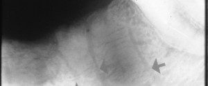
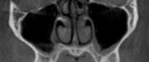
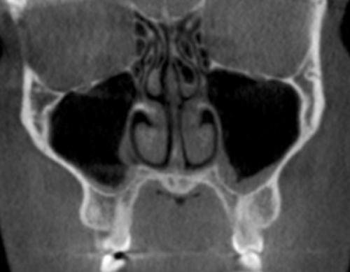
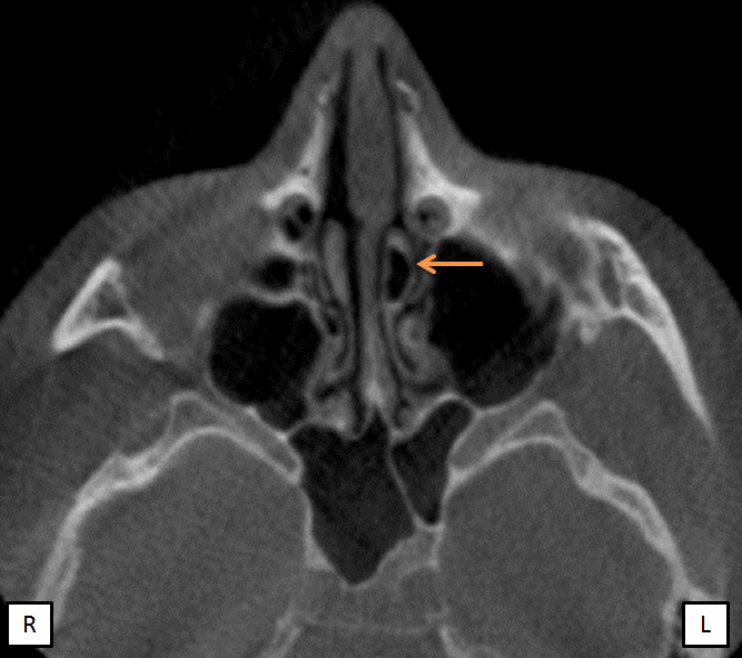
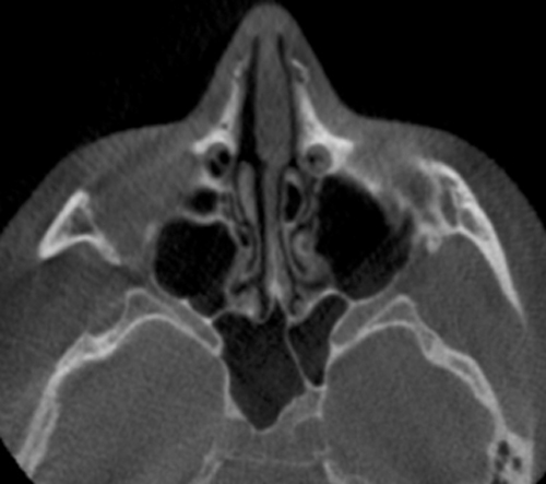
Today is concha bullosa, an aerated concha most commonly associated with the middle concha. It is seen as a well-defined radiolucent center of the concha instead of a uniform radiopaque entity.

If you have any questions or comments, please leave them below. Thanks and enjoy!
style=”display:inline-block;width:300px;height:250px”
data-ad-client=”ca-pub-8936409084392201″
data-ad-slot=”7468823574″>