This week’s anatomy is a fun one that I enjoy quizzing the students on when doing interpretations with them in the clinic, granted they may not think it’s fun when their mind goes blank on the answer. For starters when identifying the zygomatic process of the maxilla it is important to always remember to specify which bone it belongs to (MAXILLA) as there are two other zygomatic processes of the face. Ok, I digress, onto the anatomy and how it appears on radiographs.
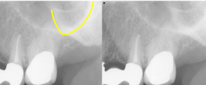
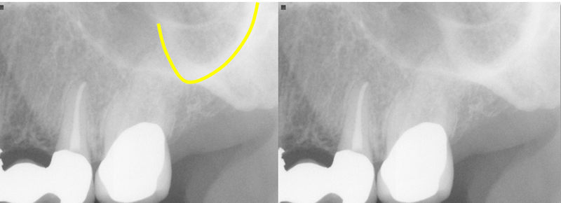
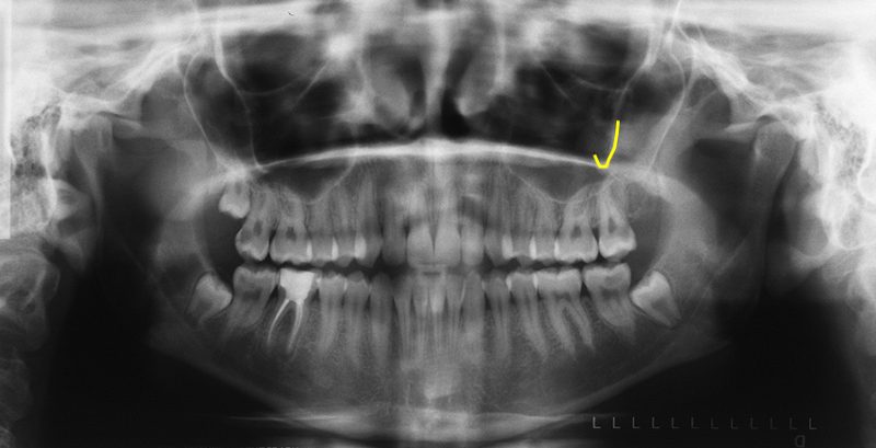
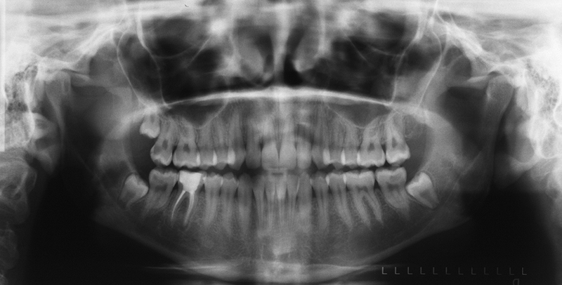
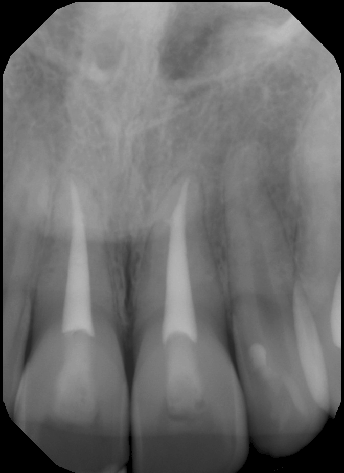
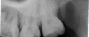
The zygomatic process of the maxilla appears as a radiopaque U, V, J shaped entity superior to the first/second molar.
If you have any questions about the zygomatic process of the maxilla, please leave them below. Thanks and enjoy!
style=”display:inline-block;width:300px;height:250px”
data-ad-client=”ca-pub-8936409084392201″
data-ad-slot=”7468823574″>
Ah! I always learn something new from your posts. Thank you
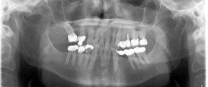
Have you done a post on the Maxillary air sinus/antrum & thier associated features on OPG?
I have not done a post on that. I will work on creating one. Keep an eye out in the next month. 🙂 Thanks for asking.
Hi, just wondering what the second radio-paque line is just posterior to the zygomatic process.
On the periapical radiographs, the zygomatic bone is seen as a radiopaque mass distal/posterior to the zygomatic process of the maxilla.