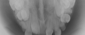
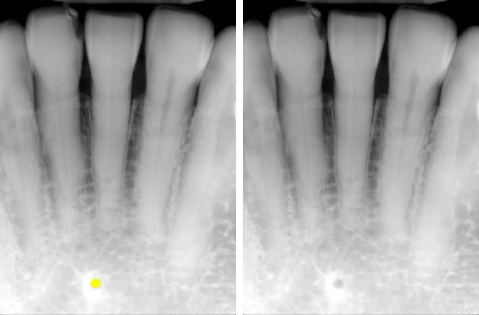
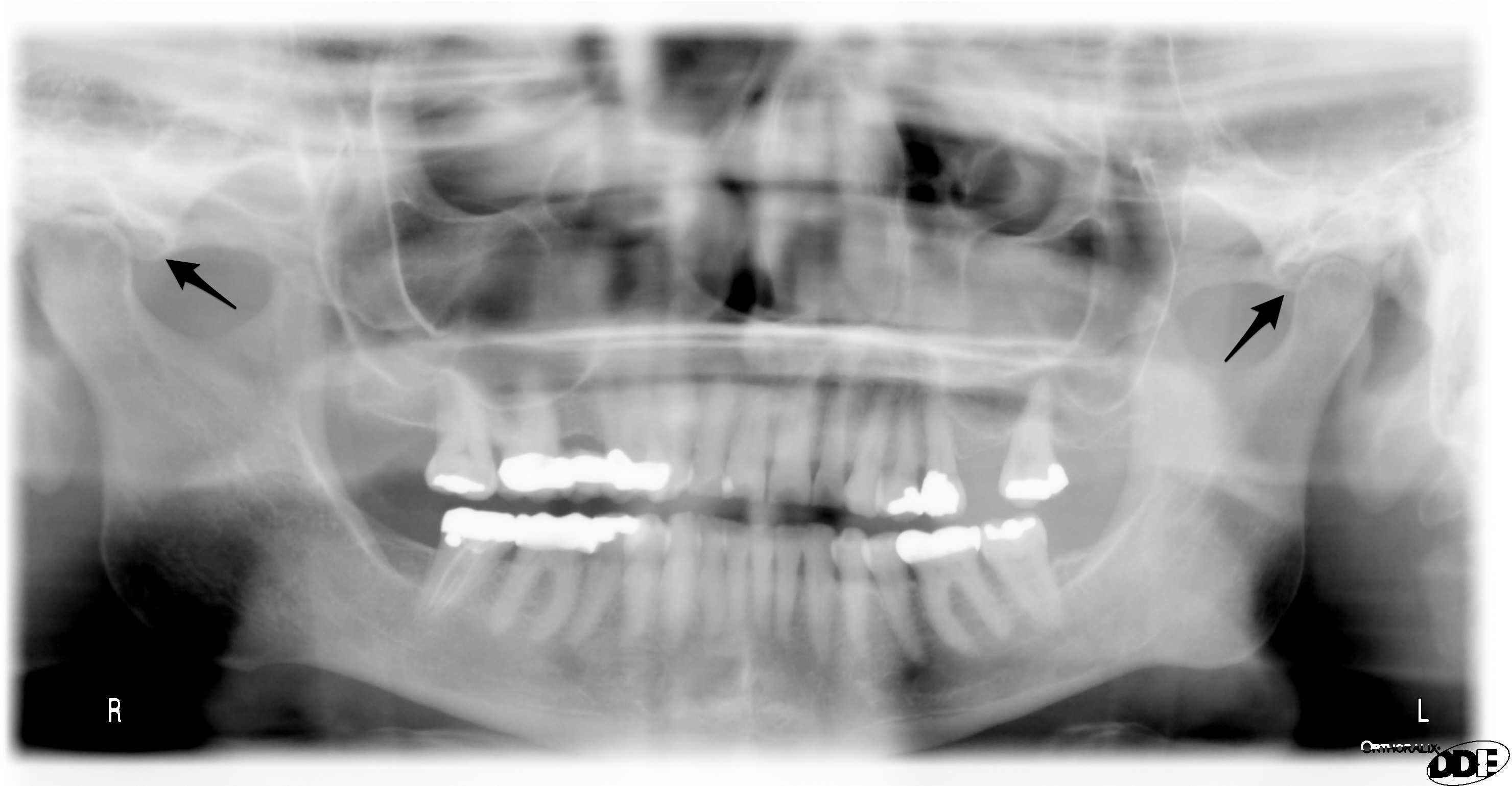
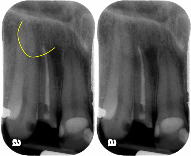
Today’s anatomy is by request for the lateral fossa also known as the incisive fossa and canine fossa. The lateral fossa is depression of the maxilla around the root of the maxillary lateral incisors. It presents as a diffuse radiolucent area around the root of the lateral incisor.
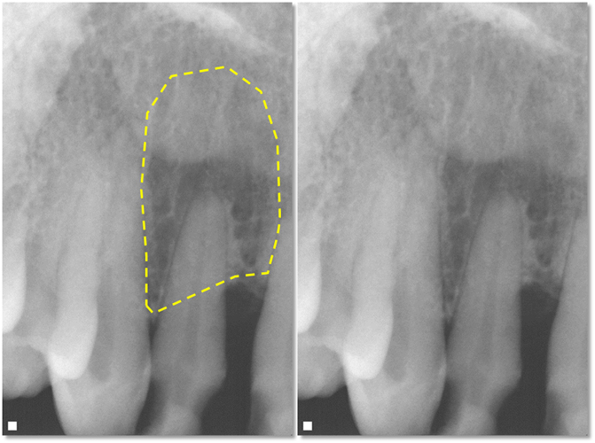
It is important to note that this area is normal anatomy and not pathosis. If there is a question of bone loss due to inflammation evaluate the lamina dura and periodontal ligament space carefully to determine if it is intact around the entire root.
If it is intact, the radiolucent is normal anatomy – lateral fossa.
If it is not intact and is continuous with the radiolucent area, pathosis of odontogenic origin should be considered.
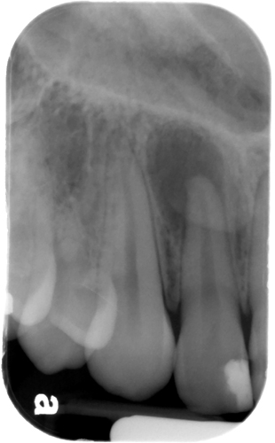
Many times the superimposition of the ala of the nose will make the lateral fossa appear more radiolucent causing over-interpretation of pathosis in this region.
If you have any questions or comments about the lateral fossa, please leave them below. Thanks and enjoy!


