This week and next I will be covering two extraoral radiographs made in dentistry. The first one is a lateral cephalometric skull radiograph which is commonly made for orthodontic purposes. Here is how to position a patient for this radiograph.
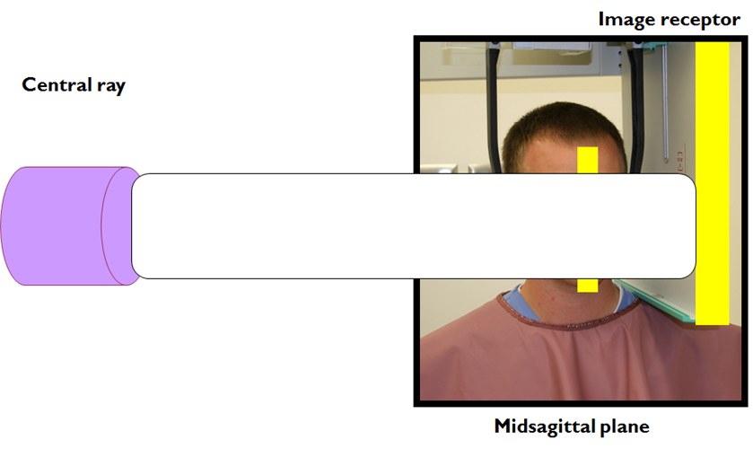
1. Place the midsagittal plane of the patient parallel with the image receptor. If there is a craniostat to help position this will be GENTLY placed in the ear canals to achieve this.
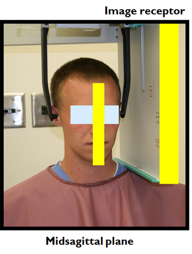 2. The source of radiation should be perpendicular (90 degrees) to the image receptor and centered over the sella turcica. (Note: newer machines have this built in so you can skip this step :)).
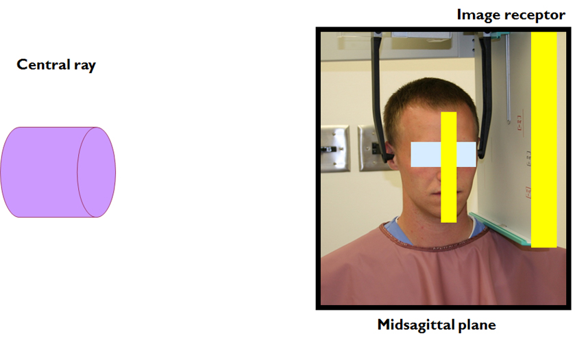 3. Irradiate the patient. (Since you can’t see x rays I’ve noted them with a white bar for the image)
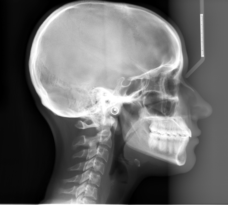 Next week: Posteroanterior (PA) skull radiograph.
Fun question – does anyone know the difference between a lateral cephalometric skull radiograph and a lateral skull radiograph?
If you have any comments or questions, please leave them below. Thanks and enjoy!
great website Dr! simple and concise.
Can I ask regarding irregularities in the cehalometric images, the diagnostic center could not explain what it is and they said they have calibrated their machine recently. May I send a copy of the xray? the irregularity is only evident on the lower anterior part of the lateral ceph image. thanks.
-JL
Thanks and yes you may send me a radiograph (no guarantees that I’ll know what the issue is though :)). My email is drgstoothpix (at) gmail (dot) com
I have to spell it out so that spambots don’t’ find it and overload my email.
Re: difference between a lateral cephalometric skull radiograph and a lateral skull radiograph? basically the same, cephalomatric skull requires the spine to be straight.
Interesting – I haven’t heard the difference in relation to the spine. I was taught that it was all based on whether the x ray unit has a craniostat to position the patient versus eyeballing it (so to speak).
What digital machine do you use to obtain your full head lateral ceph image? Thank you.
At my current institution they are using Carestream in the orthodontic department.