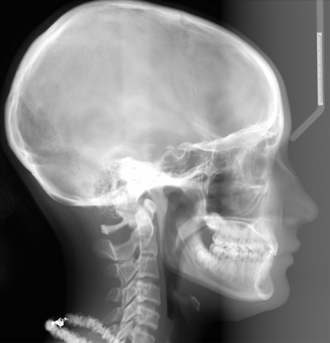
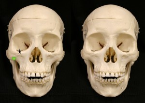
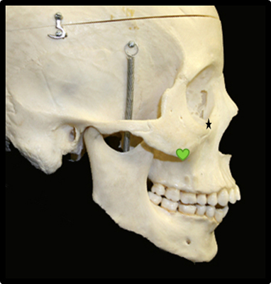
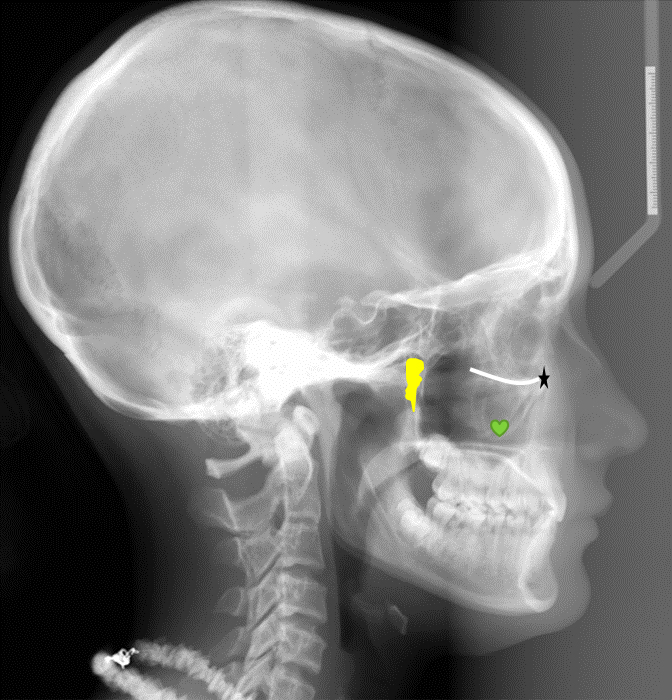
Onto part 2 of lateral cephalometric skull anatomy frequently used in orthodontics. Again identified on a dry skull first followed by the radiograph.
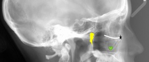
Orbitale (black star) – The lowest point of the orbital margin (also the anterior point for the Frankfort horizontal plane).
Key ridge (green heart) – The inferior portion of the zygomatic process of the maxilla.
Key ridge (green heart)
Floor of the orbital cavity (white line – directly posterior to orbitale)
Pterygomaxillary fissure (yellow inverted teardrop area)
 If you have any questions or comments, please leave them below. It appears that this will be a 7-8 part series so I’ll start posting them out twice a week starting this week. Thanks and enjoy!
Dr G – I’m a second yr dental student and just wanted to let you know that your blog blows my mind! You make radiology fun and simple to understand 🙂
-Patty
Thanks Patty,
I’m soo glad you’re finding the site useful. If you have any requests for topics or diseases, please let me know and I’ll see what I can do. Thanks. 🙂
Dr. G
Hi Dr. G, I can’t believe I’ve just now found your site — it is super helpful!
I had a question about pterygomaxillary fissures: why would you see 2 pterygomaxillary fissures on some lateral cephs? Would it be caused simply by the patient being not positioned correctly, so you are seeing both L and R ptm fissures? Or could something else make this happen?
Thank you!
Colette
Colette,
Yes, if you are seeing two it’s the right and left side most likely due to incorrect patient positioning. If you have any other questions or requests, please let me know and I’ll see what I can do.
Shawneen
Wow, thank you for such a quick reply! I’m studying for my NBDE part 2 and your site is amazing 🙂
My study group and I have been slowly making our way through radiographs from a test drive, but we came upon a few pathology slides that stumped us. On some of them, we aren’t 100% sure if it is pathology or just anatomy. I think we have “good guesses,” but we can’t confirm since there were no answers provided with these particular slides. Would you mind taking a look at the slides (there are 6) to see if we’re going in the right direction? It would definitely help having a fresh — and much more experienced — set of eyes looking at them!
Thanks again,
Colette
Thanks Colette, sure go ahead and send me the links to slides along with what you are thinking to drgstoothpix@gmail.com
Shawneen
How do we clinically evaluate the key Ridge & tonsils & adenoids
I am not familiar with the key ridge. As for adenoids and tonsils, it is a matter of evaluating the airway and general dimensions (antero-posterior width) in the area of the adenoids and tonsils. Keep in mind there are many other aspects of a patient superimposed over those areas.