1 min read 3

Anatomy Monday: Nasolabial Fold (soft tissue)

This week is another soft tissue entity that can be seen on both intraoral and extraoral radiographs. The nasolabial fold presents as a diagonal transition line. A transition line is seen as a defined line where part of the radiograph appears more radiopaque due to superimposition of soft tissue. The…
1 min read 5

Case of the Week: Ectopic Eruption & Internal Resorption

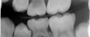
This week I have a fun case of 2 finds on 1 radiograph. This bitewing radiograph shows internal resorption (enlarged pulp chamber) of the maxillary left deciduous second molar and ectopic eruption of the mandibular left first premolar into the deciduous second molar.  Check it out below. 🙂 If you…
1 min read 0

Lateral Cephalometric Skull Anatomy – Part IV

This post will be all about the maxilla. Anterior nasal spine (yellow arrow) – anterior-most point where right and left maxilla meet on the midline forming a protruding bone spicule. A point (white arrow) – the deepest point between the anterior nasal spine and prosthion. Prosthion (yellow dotted arrow) –…
1 min read 0

Anatomy Monday: Epiglottis (soft tissue)

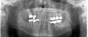
This week I wanted to show off something not commonly looked at or for on pantomographs; the epiglottis. The epiglottis will appear near the inferior aspect of the radiograph as a curved radiopaque entity. It may be superimposed over the hyoid or inferior border of the mandible.  If the airway…
1 min read 2

Case of the week: Horizontal root fracture

This week I have a case of a horizontal root fracture that is tricky to see on the initial radiograph but when another radiograph was made at a different horizontal angle it becomes clear as night and day. Take a look below. First radiograph, check out the mesial aspect of…