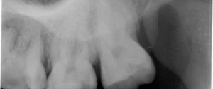
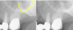
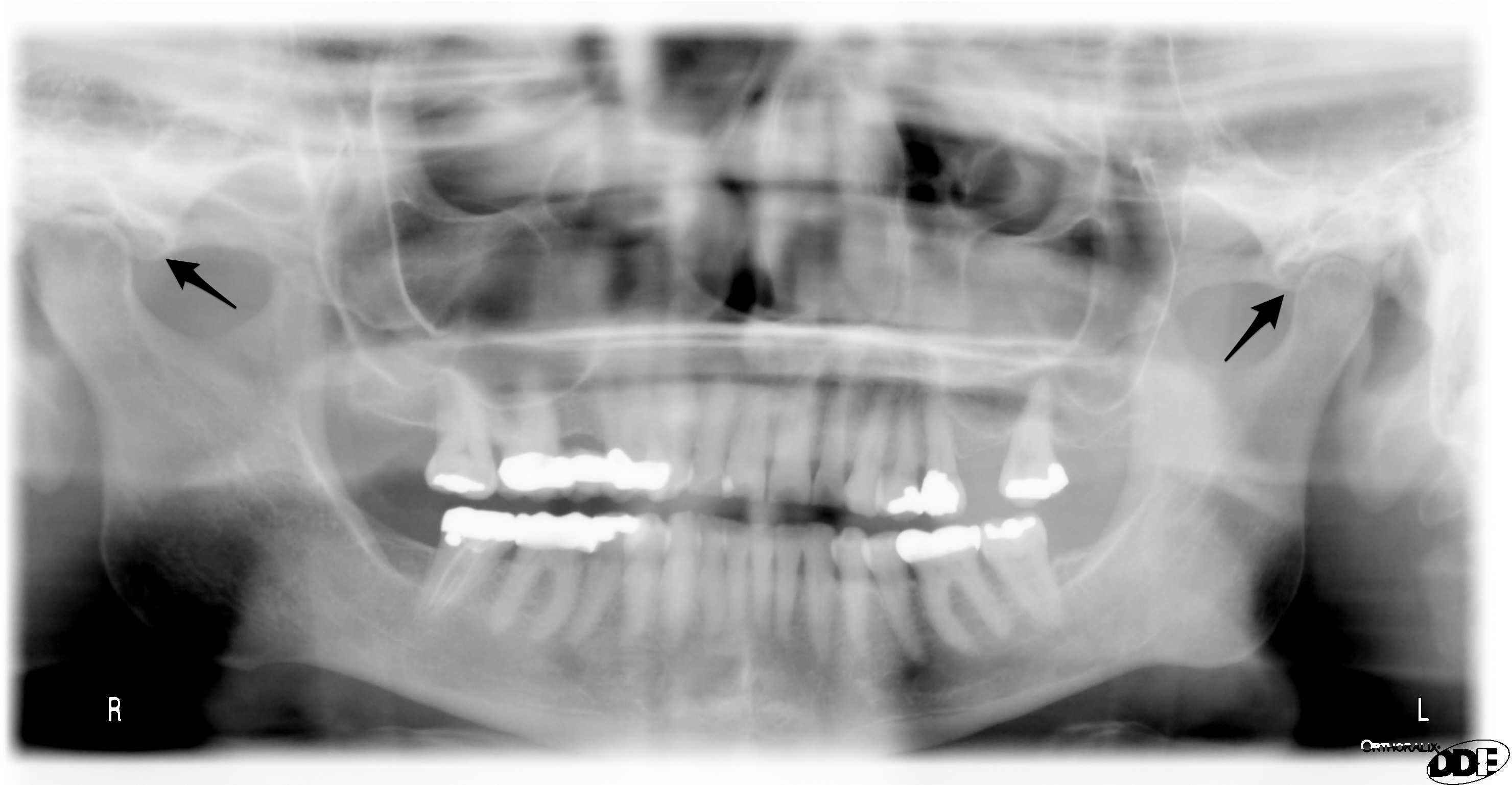
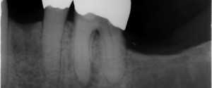
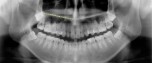
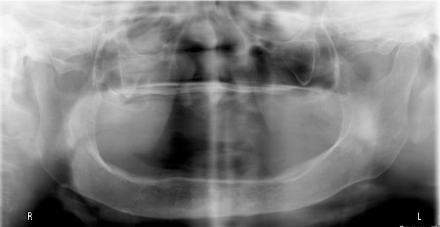
The coronoid process is the superior aspect of the anterior portion of the ramus of the mandible. It is visible on intraoral radiographs but not mandibular periapical radiographs instead it is seen on maxillary molar periapical radiographs. This typically due to the posterior placement of the image receptor and holder (XCP for example) causing the patient to open wide and move the coronoid process anterior and inferior. It will appear as a radiopaque triangle at the distal aspect of periapical radiographs. It’s real easy to spot on pantomographs as all you have to do is follow the anterior portion of the ramus superiorly. Below are some different examples and appearances of the coronoid process.
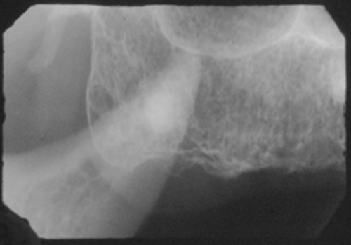
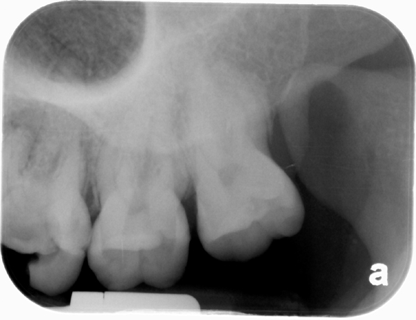
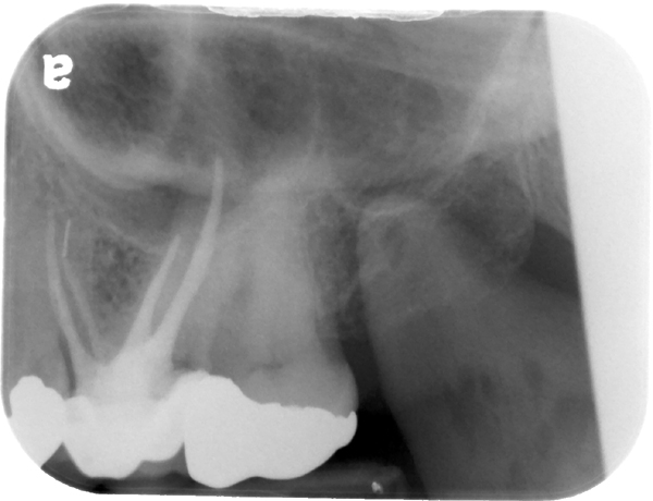
 If you have any questions or comments, please leave them below. Thanks and enjoy!
