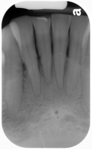
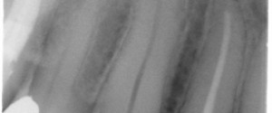
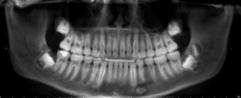
This weeks case is an interesting variant of normal anatomy – two lingual foramina seen on a periapical radiograph. Some patients can have up to three lingual foramina.
 Note the two well-defined, circular radiolucent entities inferior to the mandibular central incisors. The superior one is larger than the inferior one.
If you have any questions, comments or good examples of errors regarding these two specific criteria, please let me know. Thanks and enjoy!
SPONSOR


I have never seen this before- so thank you for sharing it. What structure passes through these foramina ? Branches of the incisive nerves and vessels?
Based on my conversations with the anatomy instructor here its merely an anterior extension of the inferior alveolar nerve. I’ll have to ask to find out if it has a specific name.
Is it reported in literature earlier
Not that I’m aware of. If you know of an article somewhere, please let me know. Thanks.