This is the second and last post on occlusal technique. For a refresher on basic occlusal technique go here.
Mandibular Occlusal Radiographs
Mandibular Anterior
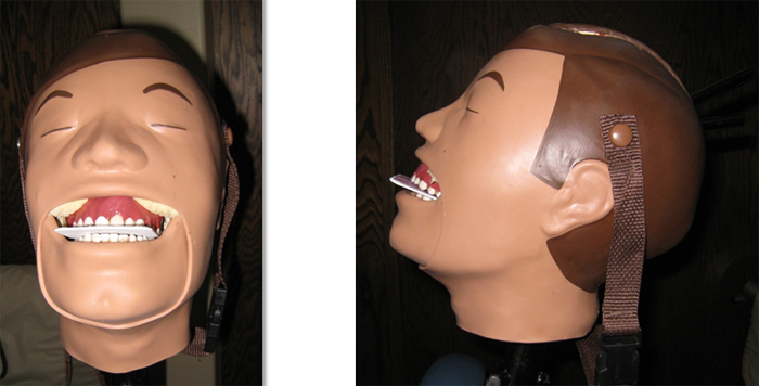
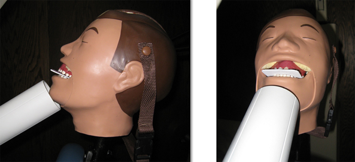
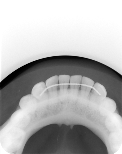
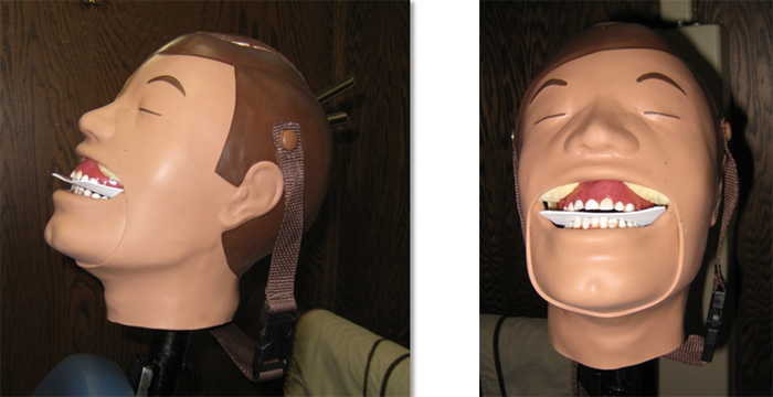
The mandibular anterior occlusal radiograph is frequently made on both adults and children. On adults, it is made with a size 4 film/phosphor plate with the long axis antero-posteriorly or horizontally. It is important to have a minimum of 1 cm of film/phosphor plate anterior to the mandibular central incisors. The central ray (PID) is aimed with a vertical angle of -55 to -60 degrees, a horizontal angle of 0 degrees and centered on the chin. For a child, a size 2 film/phosphor plate is used with the long axis laterally. The central ray is positioned the same as for an adult.
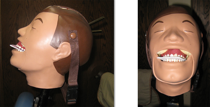
Long-axis of film placed antero-posteriorly with a minimum of 1 cm of film anterior to central incisors.
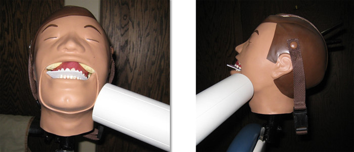
PID positioned (centered on chin) with a vertical angle of – 55 to -65 degrees and a horizontal angle of 0 degrees.
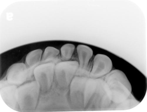
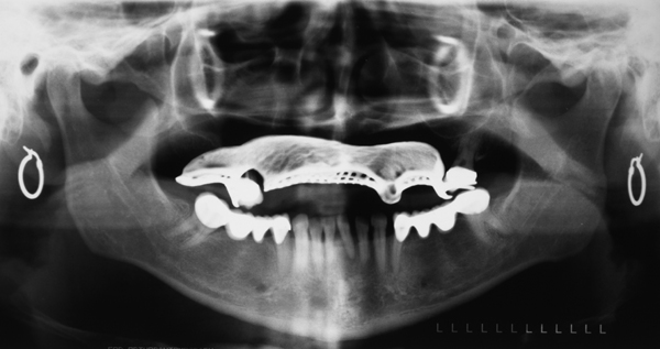
Example adult mandibular anterior occlusal radiograph.
Example child mandibular anterior occlusal radiograph.
Mandibular True (Mandibular Cross-sectional)
The mandibular true occlusal radiograph is one of the most commonly made occlusal radiographs. It is made using a size 4 film/phosphor plate with the long axis laterally. The central ray (PID) is aimed with a vertical angle of -90 degrees, a horizontal angle of 0 degrees and centered on the midline of the patient near the middle of the film/phosphor plate. The film/phosphor plate can be centered in the oral cavity or positioned to a side depending on what you want to view.
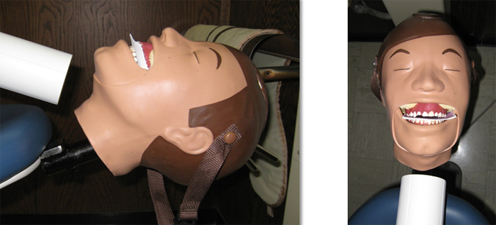
Long-axis of film placed horizontally.
PID positioned (centered on midline of patient in center of film) with a vertical angle of -90 degrees and a horizontal angle of 0 degrees. This is where DXTTR having no neck made it difficult to show a true – 90 degree vertical angle.
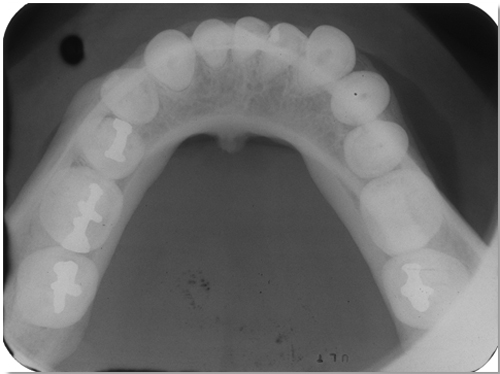
Example mandibular true occlusal radiograph.
Mandibular Lateral
The mandibular lateral occlusal radiograph is made using a size 4 film/phosphor plate. The film/phosphor plate is positioned with the long axis parallel to the facial surfaces of the posterior teeth. The central ray (PID) is aimed with a vertical angle of -50 degrees through the center of interest (typically the molar or premolar region).
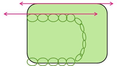 Diagram showing long-axis of film parallel with facial surfaces of posterior teeth (pink arrows).
Long-axis of film parallel with facial surfaces of posterior teeth.
PID positioned (centered on area of interest) with a vertical angle of -50 degrees and a horizontal angle of ~90 degrees.
No example mandibular lateral occlusal radiographs to share. 🙁 If anyone has one they’d be willing to share and let me add to this post, please let me know.
Thanks and enjoy!
One thought on “Radiographic Technique: Mandibular Occlusal Radiographs”
Comments are closed.