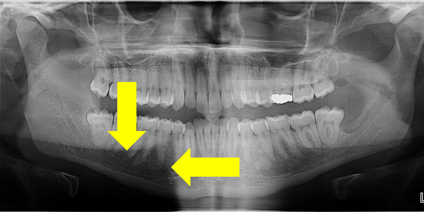
This week I wanted to show an area on pantomographs that I am frequently asked about. The area I am talking about is the radiolucent area as noted by the yellow areas inferior to the mandibular posterior teeth.
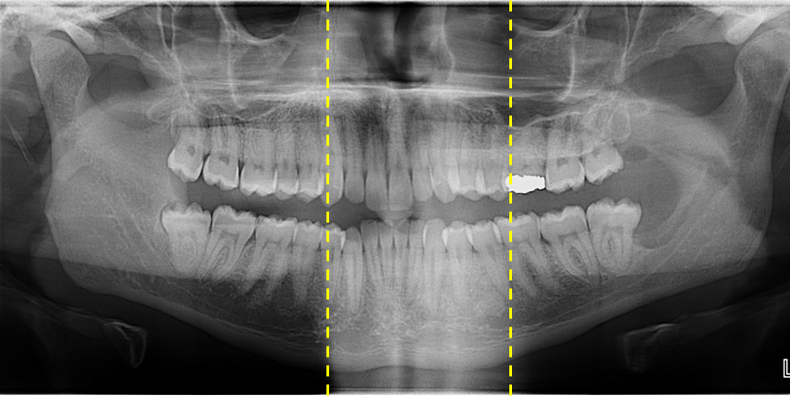
This area is typically where an observers eye are drawn due to the contrast of how radiolucent it appears compared to adjacent structures. This area is a normal projection of pantomographs and created by the superimposition of two entities and normal anatomy. The first entity that is superimposed is the cervical spine (vertical yellow dotted lines). This creates an increased radiopaque area in the mid-line of the radiograph.
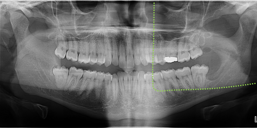
The second superimposition is a ghost image of the opposite ramus (green dotted line). This creates an increased radiopaque area superior to the mid-root and apical portions of the mandibular posterior teeth.
The last thing that is making this area appear more radiolucent is the normal anatomy of the submandibular salivary gland fossa where the mandible is thinner in this region creating an appearance of more radiolucent bone compared to the crest of the alveolar ridge.
When looking at a pantomograph in the future, if you find your eyes drawn to this area make sure to ask yourself some questions before possibly exposing your patient to excess radiation. Should you have any questions on this specific area on a pantomograph, please let me know. Thanks and enjoy!
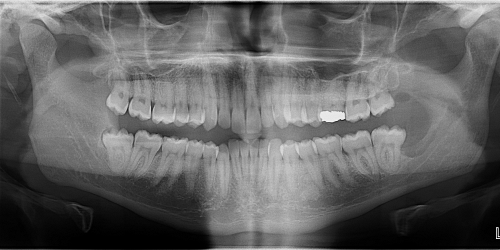
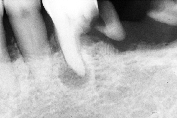
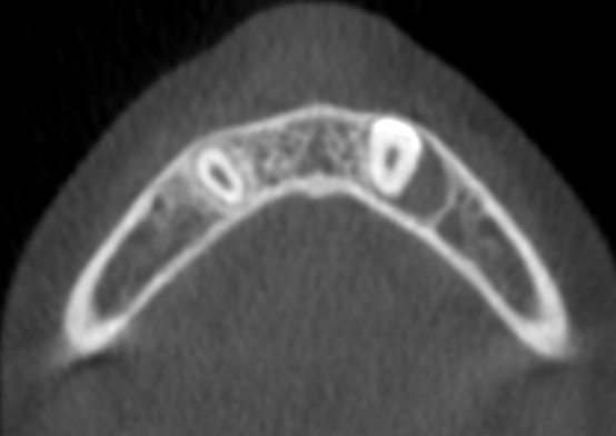
One important thing there is enostosis i.r.t 34 and 35 and bone loss can be seen in mandibular left posterior region due to extraction of third molar
Yes, on both the enostosis and bone loss due to third molar extraction.
Thanks dr for this wonderful information…can u tell me radiolucent area is seen in opg in ramus region anterior and posterior to inferior alveolar canal…is it normal?why is that?
It sounds like the area you are describing is the superimposition of the airway over the ramus making the mandible appear more radiolucent.