Ok, I am back and now onto pantomographs. Since these images are larger and will take up much more space I have decided to break them down into 2 parts. I also will have several anatomy identified on each radiograph as well. Here we go.
Mandible
*The mandibular condyle (two thin arrows pointing down) appears as a rounded radiopaque area in the glenoid fossa on the lateral aspect of the radiograph. *new anatomy not visible on intraoral radiographs.
The coronoid process (triangles) appears as a triangular radiopaque area superimposed over the maxillary tuberosity and posterior maxillary sinus.
The lingual foramen (circle) appears as a small radiolucent circle directly inferior to the central incisors.
The inferior border of the mandible (multiple thick arrows pointing up) appears as a thick radiopaque band.
The mental foramen (circle) appears as a round to oval radiolucent area seen near the apex of the second premolar.
The inferior alveolar nerve canal / mandibular canal (two curved lines) appears as radiolucent band with two thin radiopaque lines running parallel to each other (superior and inferior).
Maxilla
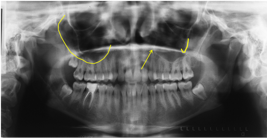
The Y line of Ennis / inverted Y (arrow) is not a true anatomical landmark but seen only on radiographs due to superimposition of the floor of the nasal cavity (straight radiopaque line) and the border of the maxillary sinus (curved radiopaque line)
The border of the maxillary sinus (curved line on right) appears as a curved, thin radiopaque line superior to the roots of the canine and posterior teeth. The maxillary sinus appears as a radiolucent area superior and medial to the border of the maxillary sinus.
The zygomatic process of the maxilla (J shaped line on left) appears as a U, V or J shaped radiopaque line. It is superior to the first and second molars.
Nasal region
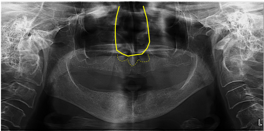
The borders of nasal cavity (solid U shaped line) appears as radiopaque lines medial to the maxillary sinuses.
The soft tissue of the nose (dotted curved line) appears as a radiopaque area superimposed over the maxillary anterior teeth. This is more commonly seen on patients who are edentulous in the anterior maxilla.
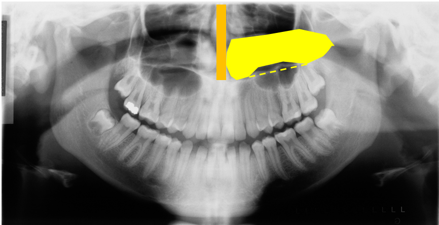
The nasal septum (orange vertical block) appears as a radiopaque band going superior from the floor of the nasal cavity. It is on the midline.
The inferior nasal concha (yellow horizontal mass) appears as a round to ovoid radiopaque mass superior to the floor of the nasal cavity.
The floor of the nasal cavity (dotted horizontal yellow line) appears as a thin straight radiopaque line.
Next = Pantomographs Part 2
I hope you find this first post on pantomograph anatomy informative. While I didn’t cover all the anatomy visible, these are the more commonly seen entities. If you have any questions, please let me know.
Enjoy!


Love the quality and description. thank you
Thanks. 🙂
Thank you for the detailed information!
The zygomatic process of the maxilla (J shaped line on left) appears as a U, V or J shaped radiopaque line. It is superior to the first and second molars.
Is this line actually showing the floor of antrum? as I read from other textbooks
Are you referring to the image left (patients right) or patients left (image right)? The J on the patients left (image right side) is the zygomatic process of the maxilla. The yellow curved line on the image left side (patients right) is the floor of the maxillary sinus or antrum is an older term for the sinus you may have heard.