I am starting a new series of posts on anatomy on radiographs. There will be two posts on intraoral radiographs (Part I – anterior and Part II – posterior), one on occlusal radiographs, one on pantomographs and lastly one on skull radiographs (primarily lateral cephalometric skull radiographs).
Anatomical radiographic appearances

Foramina – round to ovoid radiolucent entities, may or may not have a radiopaque/corticated edge.

Canals – radiolucent line or band, may or may not have radiopaque/corticated edge(s).
Now on to the anterior portion of intraoral radiographs.
Mandible
The mandible is a nice place to start as there are fewer anatomical landmarks identifiable in the anterior region (compared to the maxilla). There are four anatomical landmarks frequently identifiable: lingual foramen, nutrient canals, mental ridge, and inferior border of the mandible.

The lingual foramen appears as a small radiolucent circle directly inferior to the central incisors. It is not always visible on every patient.

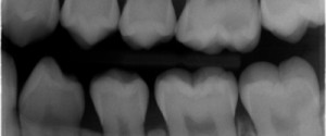
Nutrient canals (vascular canals) are canals containing blood vessels coursing throughout the maxilla and mandible. They are very small and more commonly seen in areas of thin bone (hence the anterior mandible). They appear as a radiolucent line or band coursing in a vertical direction.

The mental ridge (mental triangle) appears as two oblique thick radiopaque bands that that meet in the midline giving it the appearance of an inverted V or triangle shape. This is more commonly seen when an increased negative vertical angle is used.

The inferior border of the mandible appears as a thick radiopaque band.
Maxilla
The maxilla has quite a bit more anatomy evident including a good portion of the nasal cavity.

The intermaxillary suture appears as a thin radiolucent line between the two maxillary central incisors. It may not be visible on all patients.

The incisive foramen appears as a round to ovoid radiolucent area between the roots of the maxillary central incisors.

The anterior nasal spine appears as an inverted radiopaque triangle or V-shaped. It is on the midline and superior to the apices of the maxillary central incisors.

The incisive fossa appears as a well-localized radiolucent area around the root of the maxillary lateral incisor. This is due to a decrease in bone thickness in this region.

The soft tissue of the nose appears as a radiopaque area superimposed over the maxillary anterior teeth. The tip of the nose is seen over the maxillary central incisors. The ala of the nose is seen over the lateral incisors.

The floor of the nasal cavity appears as a thin straight radiopaque line.

The nasal cavity appears as a radiolucent area superior to the floor of the nasal cavity.

The nasal septum appears as a radiopaque band going superior from the floor of the nasal cavity. It is on the midline.

The inferior nasal concha appears as a round to ovoid radiopaque mass superior to the floor of the nasal cavity.

The Y line of Ennis (inverted Y) is not a true anatomical landmark but seen only on radiographs due to superimposition of the floor of the nasal cavity (straight radiopaque line) and the border of the maxillary sinus (curved radiopaque line).
The border of the maxillary sinus appears as a curved, thin radiopaque line superior to the roots of the canine and posterior teeth. The maxillary sinus appears as a radiolucent area superior to the border of the maxillary sinus.
I hope you find this first post on intraoral anatomy informative. If you have any questions, please let me know.
Enjoy!

Very easy & informative
Thanks. 🙂
Very informative, thanks. Do you have any presentations showing pathology?
There are some videos on radiographic pathosis on the youtube channel made by students. What type of presentation exactly were you wanting to see and possibly I can try working on creating something in the future. 🙂
it was awesome.I am a dental student and studying for NBDE part2.your images and explanation is much better than decks.
Thanks a lot for sharing information.
Thanks soo much for the comments. I’m glad that you found this post useful. If you have any topics you’d like to see, please just leave a comment and I’ll see what I can do. 🙂
Great!
Thanks. 🙂
Wow! Thank you….
Studying for my RHS exam this really help me. My brain was shutting down from information overload! Thank you for you post!
Thanks and good luck. 🙂
thanks Dr.shawneen for this part ,really it helps me alot :)….please i need explanation of premolar & molar region 🙁
What part of the premolar and molar region are you stuck on? Or do you just want a single radiograph and all the anatomy identified on that radiograph? Thanks and glad you found this helpful. 🙂
Hi ,
I found ur site to be very useful and informative.If you could post and teach how to read an opg it would be of immense help.Thanks in advance.
Thanks and I will work on your suggestion. Thanks for asking and if there are any other topics you are interested in please let me know. 🙂
Hi !
Very useful information.
Can you plz tell me how to differentiate between canine and lateral fossa on a periapical radiograph?
Thanks !
I’ll post an Anatomy Monday on that topic in the coming weeks. Thanks for asking. 🙂
Very helpful clarified what is radiopaque or translucent and great images will help me w my landmark activity and exam for radiology
Thanks and glad you find everything useful. 🙂
Excellent very helpful!
Thanks.
very helpful thanxs
You’re welcome. Glad you are finding the site useful. 🙂
Dr. G., I visited your site last year when studying for my second year oral radiology exam. Now that my third year exam is a week away, I rediscovered your site and just wanted to let you know how much it’s improved and grown. Thanks so much for keeping it up to date and continuing to be very informative! I keep forgetting but I’ll let Dr. Ernest Lam you say ‘hi’; we’re super luck to have him as one of our instructors! 🙂
Thanks. 🙂
Very useful for my revision! Thank you!
You’re welcome. 🙂
can u please help me in finding opg landmarks which most often be confused for pathologies
Working on a post for this and it will be coming out in the next few weeks. Thanks for asking.
Thanks so much for sharing!
Very informative and useful, thank you Dr!
Thanks. 🙂
Thank you so much! this help me a lot!
You’re welcome! Glad you found it helpful. 🙂
I am studying for NBDE II and I love the quality of these pictures and the description. They a very good review source. Thank you for making them available to all.
Thanks and part of the reason this site exists.
Wowww super