This is the beginning of a 4 week series going over the settings on a x ray unit and how they affect image quality. The first topic I am going to cover is kilovoltage peak or kVp. Changing the kVp on a unit is directly associated with the overall contrast of an image (referred to as the gray scale of contrast). Some x ray units have a fixed kVp (typically in the range of 65 – 75) and some are changeable (from 50 – 100). For those units with a fixed kVp you will not be able to change this setting on your x ray unit.
A quick note, changing kVp affects the QUALITY of the x rays produced. Now onto how it does that. (Now we are getting into some really technical stuff)
Increasing the kVp setting (ie, 60 to 80) increases the potential difference between the cathode and the anode in the x ray tube of the x ray unit. This increases the velocity and kinetic energy of electrons moving from the cathode to the anode (target). This results in x ray photons produced with increased kinetic energy. This increased kinetic energy allows the x rays to have more penetrability of objects. (ie, more of the x rays will pass through the head and neck region to the film/sensor.) While this sounds like a great idea, the problem is that as you continue to increase the kVp the image quality degrades by creating an overall grayer image with little contrast.
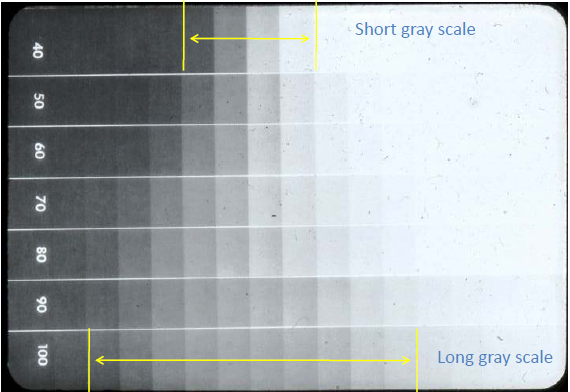
A small kVp (ie, 40 in the diagram below) will produce an image of high contrast (note the visible difference between the boxes). The problem is that few grays are visible. This is referred to as a short gray scale. A high kVp (ie, 100 on the diagram below) will produce an image with low contrast (note the overall gray line with few blacks and whites). This will show many different grays. This is referred to as a long gray scale. The problem is that the grays are so similar in shade that it is difficult to detect small changes between them creating a problem when interpreting radiographs for small changes (such as incipient carious lesions).
Since the extremes have both good and bad about them, in dentistry we have split the difference (so to speak). Ideal kVp settings typically are between 65 – 75 (what most fixed units have). This creates an image with more grays than at 40, but with more visible difference between those gray shades than at 100. See the diagram below to see the difference between the different kVp settings.
I hope these few points about kVp setting changes are informative to you and give you a better understanding of the how this setting affects image contrast. If you have any questions or comments about kVp, please let me know.
Thanks and enjoy!


why low contrast is desirable in dental radiography, as more gray shades will cause confusion and make diagnosis more difficult?
A low contrast such as with 90 and 100 kVp is not desirable in dentistry as yes, it would make interpretation of small changes much more difficult. We aim for 60 to 70 kVp making the differences between the black/gray/white shades evident on the final image. Please let me know if this helps or you have any more questions. Thanks. 🙂
To detect a root fracture radiographically would you want a shorter gray scale (ie: more contrast, lower kVp)??
Changing the kVp can help in some cases but what is more important would be changing the horizontal and vertical angles and taking another radiograph. Interesting question, thanks for asking. 🙂
Dear Dr. Shawneen Gonzalez, I am not a radiologist so forgive my question if it appears trivial. If I am not wrong, the ability of the x-ray photons to penetrate a body (decreases) increases with (decreasing) increasing kVp. Consequently, at a (lower) higher kVp I expect an x-ray radiogram appears (brighter) darker, resulting in a (larger) shorter gray scale. This is in contrast with the showed picture. Where am I wrong?
Luca,
You are correct that an increased kVp leads to an increased penetration of x rays through the body. You are also correct that a higher kVp leads to a darker radiograph. It’s very last part that is off regarding the gray scale. Due to more x rays (high kVp) passing through the body more pass through various objects and interact with the image receptor creating a longer gray scale. A low kVp does not have the power for many of the x rays to pass through different objects hence the shorter gray scale. Does this help or make it more confusing? Thanks. 🙂
Thanks for the information on your site. Pls sir, I need help regarding the equivalence of kGy and kVp. that is how can one convert from kGy to kVp or vice -versa. PLEASE HELP.
Thank U.
I’m a little confused as to exactly what you are asking as Gy is a term to described dose and kV refers to electrical aspects. How was it presented to you? Thanks.
Im a radiography student currently, just to be sure….short scale= high kVp, less greys and long scale=low kVp, more greys
short grayscale = low kVp (more blacks and whites)
long grayscale = high kVp (overall more gray image)
Thank you for this post, Dr. Gonzalez. I have a question regarding how to adjust kVp for the detection of root fractures on an xray. If increasing kVp lowers the contrast of an image (creating a larger gray scale), would this be helpful in the detection of the fracture line? Or would you need more contrast and therefore a lower kVp to better see it?
A lower kVp will increase contrast but the bigger issue with seeing a fracture line is dependent on the direction of x rays and the fracture plane. Two images with different horizontal and vertical angles may help with detection of a fracture more than changing kVp.
Aw, this was an extremely nice post. Finding the time and actual effort to generate a really good article… but what can I say… I put things
off a whole lot and don’t manage to get anything done.