What is vertical angle?
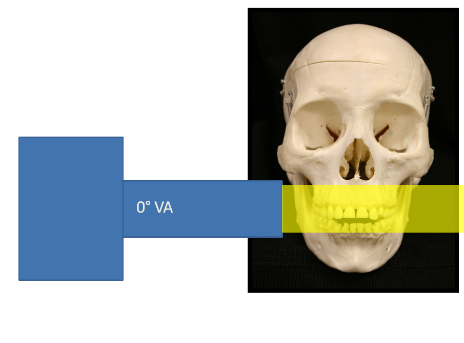
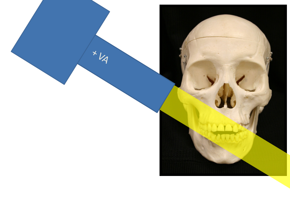
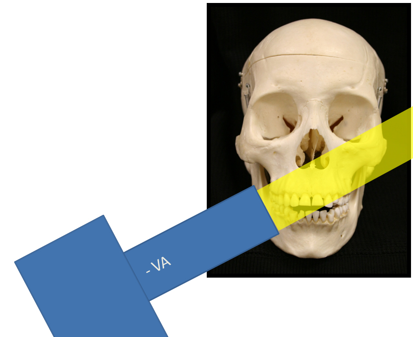
Vertical angle refers to the position the Position Indicating Device (PID or cone) is along the coronal and/or sagittal planes. A zero degree vertical angle is when the PID is positioned parallel with the axial plane. A positive vertical angle is when the source of radiation is superior to the PID and the x rays are emitted towards the floor. A negative vertical angle is when the source of radiation is inferior to the PID and the x rays are emitted towards the ceiling. To complicate things a little more, a positive vertical angle can be increased (closer to 90 degrees) or decreased (closer to 0 degrees). A negative vertical angle can also be increased (closer to -90 degrees) or decreased (closer to 0 degrees). Below are images with a very cooperative patient again (Bob) demonstrating this.
Above: Vertical angle (at 0 degrees) parallel with the axial plane.
Above: Positive vertical angle with the source of radiation superior to the PID and the x rays emitted towards the floor.
Above: Negative vertical angle with source of radiation inferior to PID and the x rays emitted towards the ceiling.
Common errors due to incorrect vertical angle
Errors due to incorrect vertical angle manifest as size changes in the image of teeth on radiographs. The different size changes are an image where the teeth appear shorter, foreshortening, or where the teeth appear longer, elongation, compared to the actual teeth in the mouth. Both of these errors occur when the vertical angle is not directed perpendicular to the long axis of the teeth being imaged. When the vertical angle is too steep (close to a positive or negative 90 degrees) this will result in a shorter image of the teeth. The diagram below shows this.
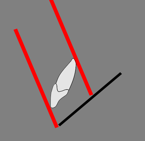 Note the red lines indicating the direction of the x rays are not perpendicular to the long axis of the tooth.
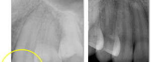
 An example radiograph of foreshortening, where the roots appear to be the same length as the crown.
When the vertical angle is too shallow (close to 0 degrees) this will result in a longer image of the teeth. The diagram below shows this.
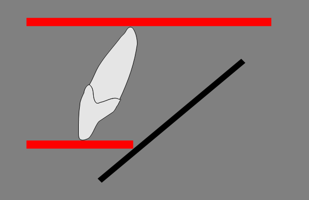 Note the red lines indicating the direction of the x rays are not perpendicular to the long axis of the tooth.
 An example radiograph of elongation.
More tidbits about vertical angle
Vertical angle errors are very important when evaluating a tooth for endodontic therapy. If the image is distorted with a shorter or longer image of the tooth, it can be difficult to determine obturation length in relation to the apex of a tooth. We use the XCP (eXtension Cone Paralleling – RINN) instruments at the school. By using these instruments or similar instruments it determines the vertical and horizontal angles based on placement of the film or sensor in the mouth. These make foreshortening or elongation less likely.
I hope this brief introduction to vertical angle is helpful in your practice or as a refresher. Please let me know if you have questions.
Thanks and enjoy!
Dr. Gonzalez!! Thank you so much for all your efforts and sharing your wealth of information. I am a dental assisting instructor and your website has been very helpful for myself as well as my students! You are awesome. And thank you again.
Thanks for the kind comments. I’m pleased to hear you are able to use this site for your students and they find it helpful. 🙂