1 min read 0

Anatomy Monday: Zygomatic Process of the Maxilla

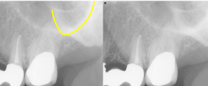
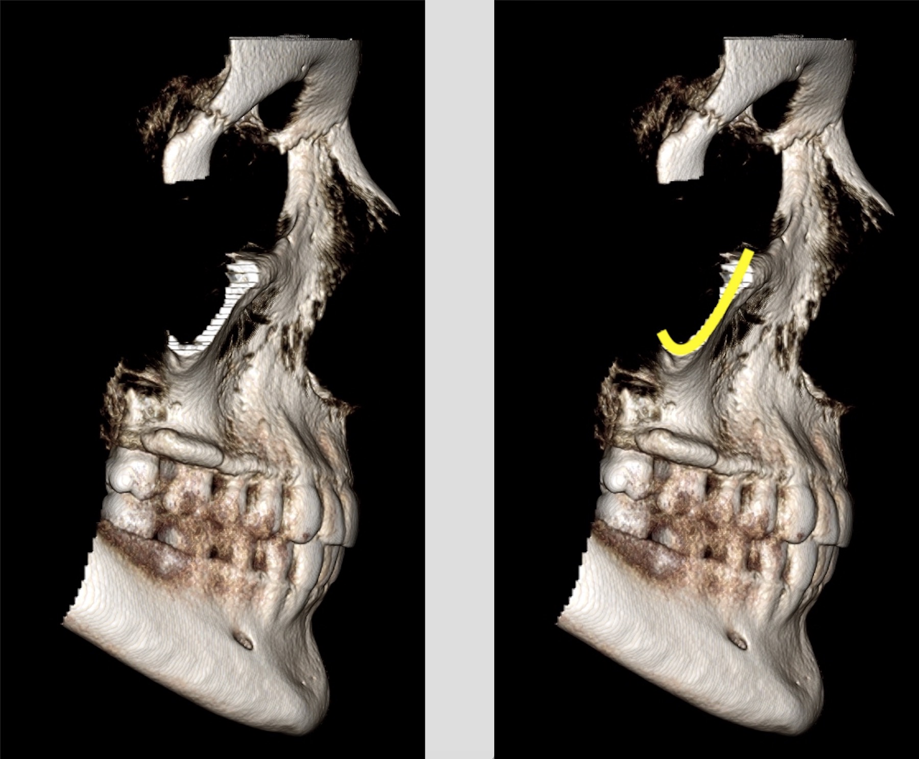
This anatomy can be tricky for some to visualize as its appearance is understood by knowing the direction of x rays and how they course through this process. So to try and show that I’m adding some 3D renderings from a CBCT scan. The zygomatic process of the maxilla appears…
1 min read 5

Anatomy Monday: Zygomatic Process of the Maxilla

This week’s anatomy is a fun one that I enjoy quizzing the students on when doing interpretations with them in the clinic, granted they may not think it’s fun when their mind goes blank on the answer. For starters when identifying the zygomatic process of the maxilla it is important…
2 min read 0

Anatomy on Radiographs: Occlusal Radiographs

This is the last post I have planned for anatomy and it will be on occlusal radiographs. Mandible Short and sweet here with only one anatomical landmark readily visible. The genial tubercles (yellow arrow) appear as a small radiopaque mass(es) extending off the lingual aspect of the mandible. Maxilla The nasal…
3 min read 5

Anatomy on Radiographs: Pantomographs Part 1

Ok, I am back and now onto pantomographs. Since these images are larger and will take up much more space I have decided to break them down into 2 parts.  I also will have several anatomy identified on each radiograph as well.  Here we go. Mandible *The mandibular condyle (two…
2 min read 7

Anatomy on Radiographs: Intraoral Radiographs Part 2

This is part 2 (posterior) of anatomy on intraoral radiographs. Mandible The mental foramen  appears as a round to oval radiolucent area near the apex of the second premolar. The inferior alveolar nerve canal (mandibular canal) appears as radiolucent band with two thin radiopaque lines running parallel to each other…